Ultrasound Case Study
Acromioclavicular Joint Osteolysis
His general health was otherwise good, and he was not taking any regular medication.
On examination, he presented with pain at the end of range of flexion and abduction of the glenohumeral joint. Scarf test was positive. There was no capsular restriction. There was localised tenderness at the acromioclavicular joint reproducing his symptoms. From the clinical examination , it was quite clear that this was a localised ACJ complaint.
On office based ultrasound we found the following image. You can clearly see the cortical irregularity on the distal clavicle (right side of the image) whilst this longitudinal musculoskeletal ultrasound scan is performed.
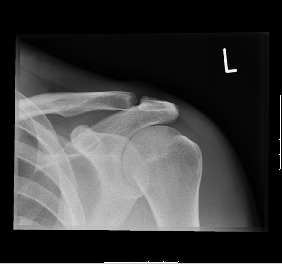
The patient was subsequently referred for an x-ray to clarify the nature of the cortical irregularity. The report is as follows and the image below ‘Acromiohumeral distance preserved. Minimal fragmentation of the lateral clavicle at the ACJ. This may be due to infection or previous trauma. Clinical correlation is advised.‘
With correlation of the X-ray findings and the clinical presentation, it was felt that this patient presented with a chronic inflammatory response to joint loading/overuse. This is sometimes referred to as Acromioclavicular Joint Osteolysis or Distal Clavicular Osteolysis. With education and a period of rest from upper body training, this patients symptoms have started to settle significantly. The access to musculoskeletal ultrasound in this case in clinic, helped assess the ACJ in greater detail and gain an appreciation of the bony changes that were occurring. This was very useful information to help the patient understand the nature of the condition and the importance of rest/modification of their activities.


Great images, I had seen those cases with MRI and x ray but not with ultrasound. Good job.
Thanks Adan, really appreciate your feedback!