Ultrasound Case Study
Common extensor tendon tear
Stuart Wildman, Extended Scope Physiotherapist
This patient in his late 20’s presented with a six month history of lateral elbow pain. This was insidious in onset, but was gradually worsening and limiting his work as a plumber. He had been referred by his GP for a further review of his elbow pain, presumed tennis elbow and for consideration of injection.
There was full range of elbow movement without complaint. There was point tenderness on examination over the common extensor tendon insertion on the lateral epicondyle. Resisted 3rd metacarpal extension was uncomfortable (2nd metacarpal extension unremarkable) along with gripping and resisted wrist extension. Potentially suggestive of a more focal complaint with the extensor carpi radialis brevis component of the common extensor insertion. Overall, the presentation was of tennis elbow, and would fit with the onset of the patients complaint.
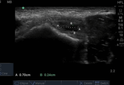
On ultrasound, there was no effusion in the anterior or posterior recess of the elbow joint. The common flexor tendon appeared intact. There was no irregularity of the radio humeral joint line. On scanning the common extensor tendon in a longitudinal view, there initially did not appear to be any irregularities to the tendon structure. But I then noticed a focal hypoechoic region within the tendon bulk in the deeper fibres consisitent with a potential common extensor tendon tear. The tendon quality was also not great, demonstrated by a lack of echogenicity. The hypoechoic defect was also clearly visible on a transverse view, although this is not often utilised as often as a longitudinal view. It is often a rule of thumb that tears should be confirmed both in a longitudinal and transverse view.
A detailed paper on lateral epicondylitis/tennis elbow and sonography can be found here by Connell et al (2001). This article provides a useful overview of the common findings on ultrasound. Interestingly, Connell et al (2001) comment that the deeper fibres of the tendon that have an articular attachment tend to be from the extensor carpi radialis brevis tendon. In this case, that would correlate with the 3rd metacarpal extension being uncomfortable.
It is always useful to reflect how integrating diagnostic ultrasound into clinical practice has informed patient management. The use of ultrasound in this case has enhanced the amount of information available from the initial consultation. Providing information on tendon integrity, and quality. It has not directly altered the management but has provided useful information that has influenced the choice of treatments. On reflection, I did not expect a patient of this age to have a partial substance tear, and it certainly makes me question the list of differential diagnoses that I often refer to when assessing the lateral elbow. I personally, do not administer many tennis elbow injections as I am not convinced of their clinical effectiveness over and above Physiotherapy in the majority of cases. However, I would also be further reluctant in this case as the patient is young, involved in manual work and he has a partial thickness tear. Further food for thought can be found in this attached article by Smith et al,prompting thought on when to inject and also the potential benefits of ultrasound guidance. This patient will continue to be managed conservatively without the use of injection. Further discussion on injections is outside of the realms of this case study.


0 Comments