Ultrasound Case Study
Knee joint effusion on ultrasound…interpret for clinical insights!
Clinical examination and history
A patient was recently referred for evaluation of the integrity of the extensor mechanism of the knee – patella and quads tendon, and to assess if there had been a rupture following a traumatic injury two days earlier. The mechanism of injury was a fall from a wall, with a twisting flexion injury. They reported immediate swelling and difficulty weight bearing. There was no audible pop. Range of movement was extremely limited due to pain. X-rays had been performed but were not conclusive. Pain inhibited muscle testing. No obvious , palpable step at the quads insertion.
Ultrasound findings and associated imaging
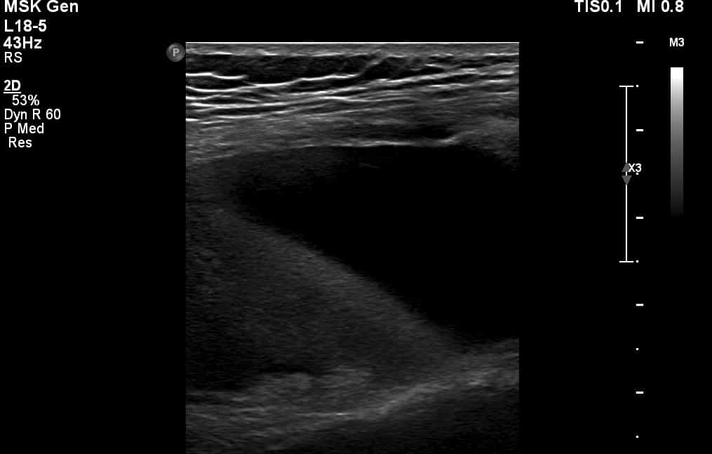
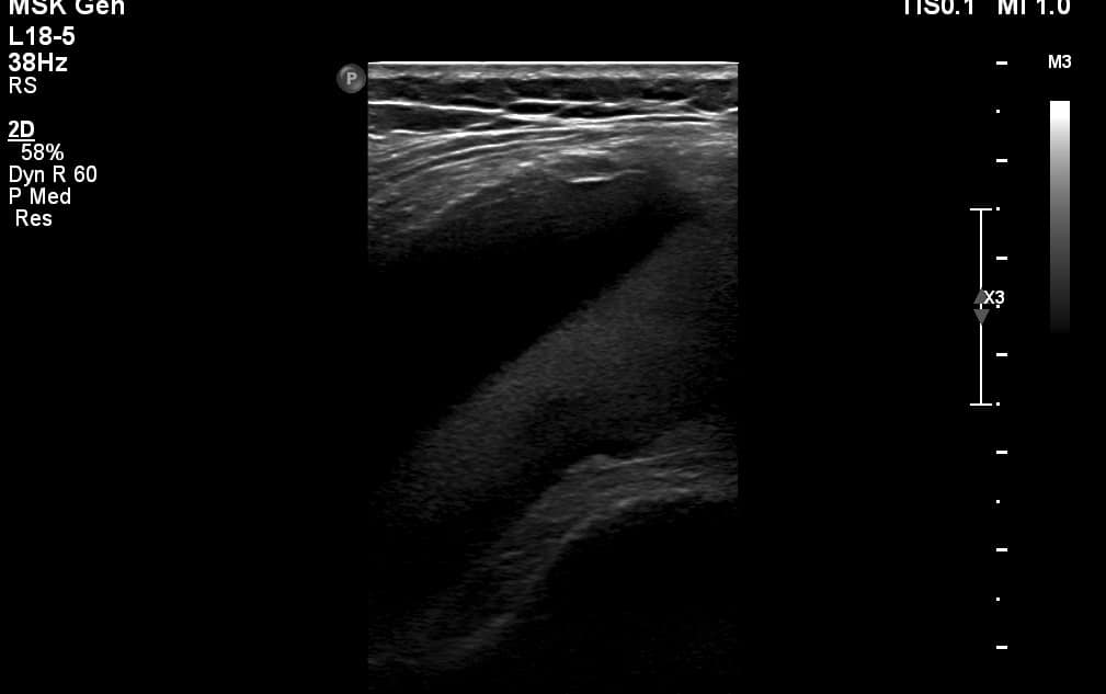
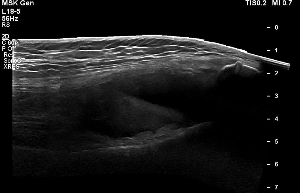
Range of movement was severely restricted by pain, therefore only limited amount of flexion could be achieved. The extensor mechanism was intact, but of clinical interest was that the ultrasound demonstrated an interesting appearance of the suprapatellar effusion which serves as a useful learning point for all of us using point of care ultrasound to assess acute, traumatic knee injuries. The effusion demonstrated a distinct line (Figure 1-3) , dividing a more hypoechoic region from a more anechoic region consistent with a Lipohaemarthsosis, where the fat and blood released from a fracture have a different sonographic appearance.
Ultrasound report ‘ The patella and quadriceps tendons were intact. There was a visible lipohaemarthosis within the suprapatellar recess. Further imaging would be advised to evaluate for a fracture.’
A subsequent CT of the knee highlighted’ There is a large lipohaemarthrosis. The patella is partially subluxed laterally and there is a large defect of the medial aspect of the patella with a corresponding defect in the lateral aspect of the distal femoral condyle. There are bony
fragments within the tissues adjacent to the patella.’
Background
Lipohemarthrosis results from the extrusion of fat and blood from bone marrow into the joint space after an intraarticular fracture. 97% of patients with an intraarticular knee fracture have a Lipohaemarthrosis (Bonnefoy et al, 2006).
Learning points from this case
- Sometimes we can become over focussed on specific structures – evaluation of sonographic appearances of fluid can give us clinical clues!
- Altering the depth of an image is important to evaluate the area in its entirety – ensure you can see the full recess or effusion to evaluate it thoroughly.
Further reading
Bianchi et al (1995) Sonographic Evaluation of Lipohemarthrosis:Clinical and In Vitro Study, Journal of Ultrasnd in Medicine, 14:279-282.
Bonnefoy et al, 2006) Acute knee trauma: role of ultrasound. European Radiology,16(11):2542-8.


Thank you. An interesting case.