Ultrasound Case Study
Posterior elbow impingement
Stuart Wildman, Extended Scope Physiotherapist and MSK Sonographer
This patient in his 30’s presented with posterior elbow impingement. He recalled a five month history of posterior left elbow pain and stiffness. He could recall a history of trauma to the elbow when he was very young, but no specific details.
His pain was focussed to the posterior aspect of the elbow. On examination he presented with a fixed flexion deformity of the left elbow, lacking approximately 30 degrees of extension. He had full elbow flexion with only slight discomfort at the end of range. Pronation and supination were preserved.
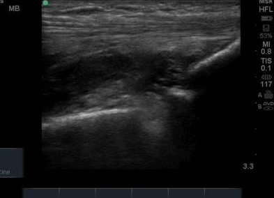
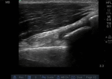
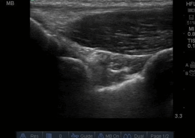
On ultrasound, there was a cortical irregularity of the distal humerus, which appeared to have a halo of fluid surrounding. This was slightly unusual and not consistent with the normal cortical outline of the posterior recess that I had encountered before. With a knowledge of the articular surfaces this would appear to cause some impingement during joint motion.
On dynamic ultrasound scanning of the elbow during flexion/extension (not easy to maintain the appropriate view!) it was possible to see the impingement of the distal humerus on the olecranon process during elbow extension and some bulging of anechoic fluid as they approximate.
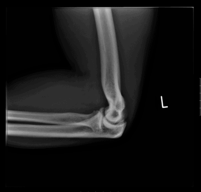
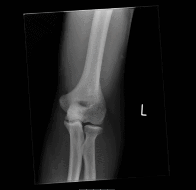
Given the significant loss of range of movement, and the subtle effusion in the posterior recess the patient was referred for an MRI to clarify the presence of any loose bodies or intra-articular cause. At this point, the previous x-rays also became available with the report indicating, ‘Modelling deformity of the distal humerus suggestive of previous trauma. No acute abnormality. Degenerative change medial humeroulnar joint.’
The MRI result indicated ‘ There is mild elbow osteoarthritis with associated synovitis. No definite loose bodies seen. The common flexor and extensor tendons are normal. Normal biceps and triceps tendons.’
This patient presents with a posterior elbow impingement, demonstrated on dynamic use of musculoskeletal ultrasound. This shows the benefit of utilising ultrasound as part of a clinical examination and integrating it dynamically. Ultrasound enabled the significance of the x-ray findings to be established.This patient has now been referred to Orthopaedics for further assessment.
Can you share a video of dynamic scan?