Ultrasound Case Study
Tibialis anterior tenosynovitis and tendinopathy
Clinical examination and history
This patient was referred for an ultrasound to further evaluate a lump on the anterior aspect of the ankle. The patient was in their 60s. On examination there was a well localised, soft lump on the anterior aspect he ankle that was not particularly tender to touch. It had been there several months and became progressively sore with walking during the day. There was no pain at rest.

Tibialis anterior anatomy on the anterior ankle, demonstrating the tendon sheath in blue.
Ultrasound imaging (further images to be added in future)
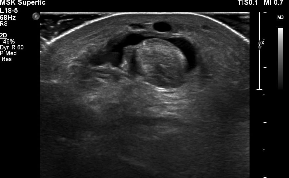
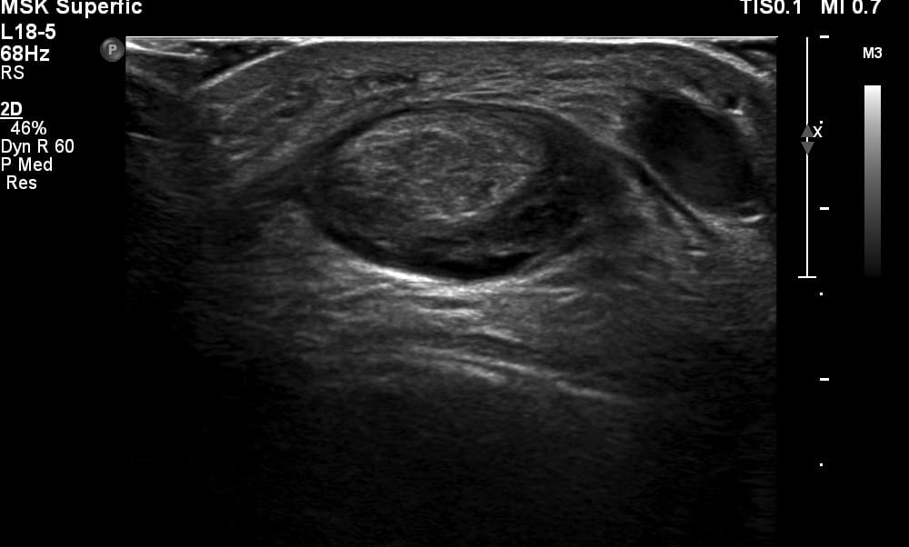
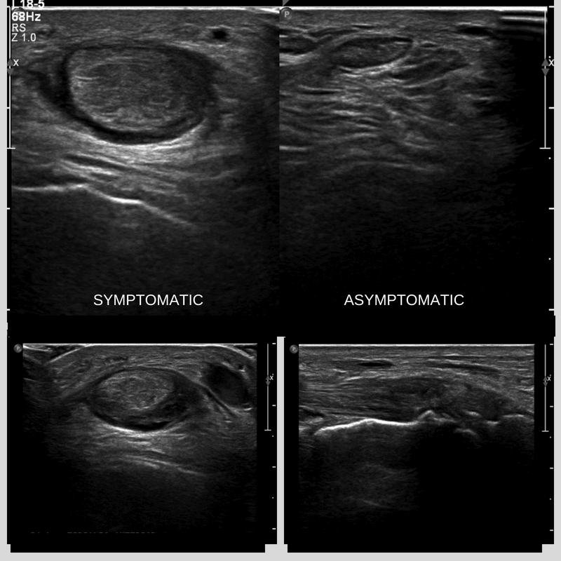
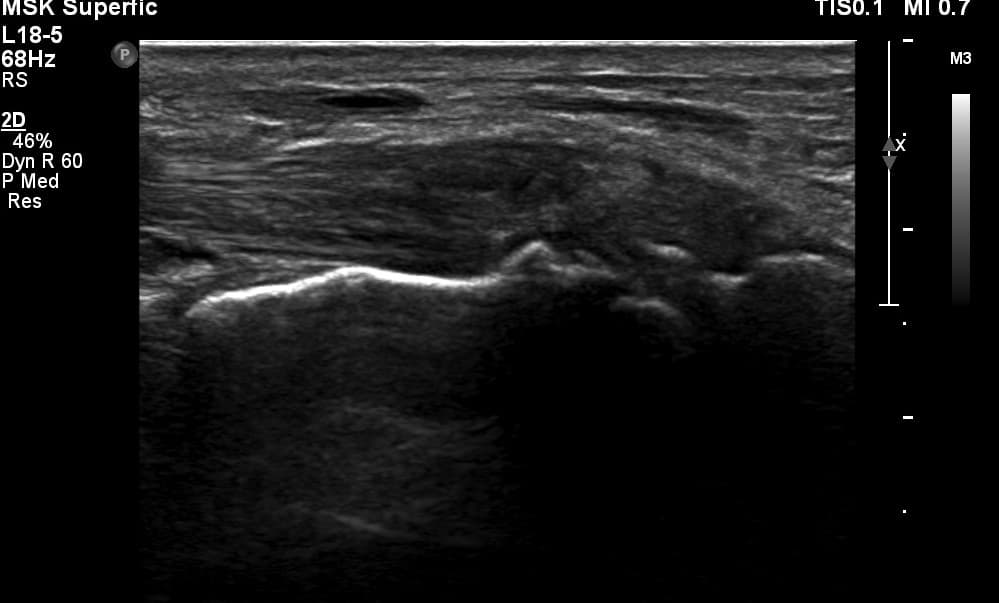
The ultrasound image encountered is demonstrated showing marked thickening of the tibialis anterior tendon short axis (Figure 1,2 and 3) with a distinct anechoic fluid collection within the tendon sheath, there was visible thickening superficial to the talonavicular joint. There was hyperaemia in the synovial thickening of the tendon sheath, unfortunately images of this were not available. It is good practice to always follow a structure in its entirety and not to be satisfied obtaining one view. The distal tibialis anterior tendon was markedly tendinopathic (Figure 4) and appeared to receiving friction from the degenerative changes at the articulation of the medial cuneiform and navicular. Often the medial cuneiform component of the distal insertion of the tibialis anterior tendon is affected (Varghese & Bianchi, 2014).
Ultrasound report ‘The tibialis anterior tendon demonstrates tendinopathy and tenosynovitis. The tendon structure appears thickened and hypoechoic, with synovial thickening and hyperaemia of the surrounding sheath. There is tendinopathy of the distal medial cuneiform insertion. There is also a fluid collection in the tendon sheath anterior to the ankle recess which correlates with the apparent lump and the focus of the referral question’
Anatomy of the Tibialis Anterior tendon
The tibialis anterior tendon is a large tendon on the anteromedial aspect of the ankle. It s a useful landmark to use as a reference point when visualising the anterior ankle region on ultrasound. The tendon forms at the level of the junction between the middle and lower thirds of the tibia, and inserts distally onto the medial cuneiform and the base of the first metatarsal bone. It is held in position partially by the extensor retinaculum.
Learning points
- Don’t assume anterior ankle pain is not related to tendon issues. Its less common but can occur.
- Be aware that the tibialis anterior tendon has a tendon sheath!
- Ensure you follow the tendon all the way to the distal insertions and can comment on its integrity throughout.
- Ensure you evaluate the dorsal recess of the midtarsal joints such as the talonavicular and navicular-cuneiform.
Useful references for further reading


0 Comments