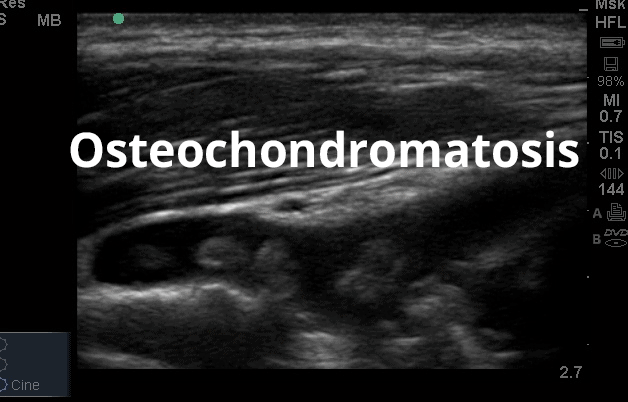
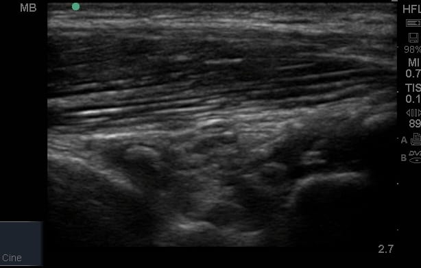
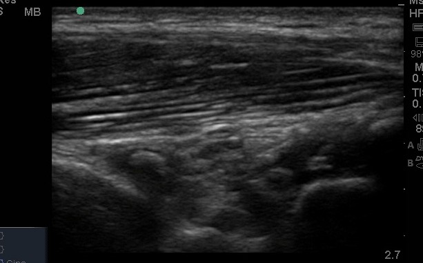
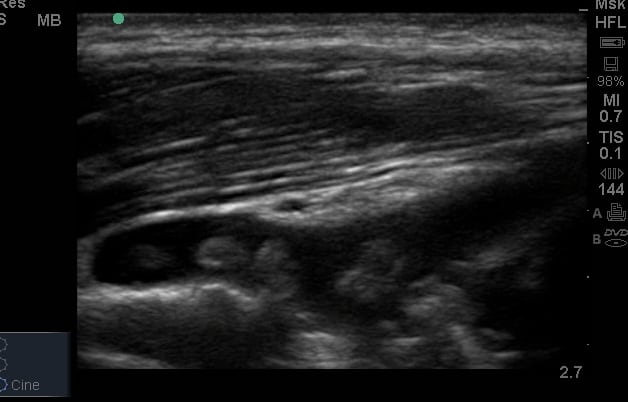
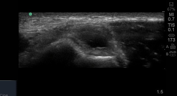
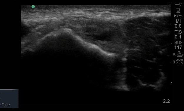
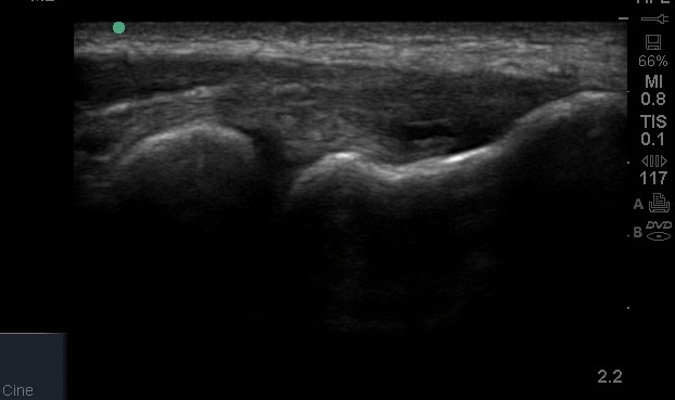
Elbow ultrasound
What structures can we see at the Elbow?
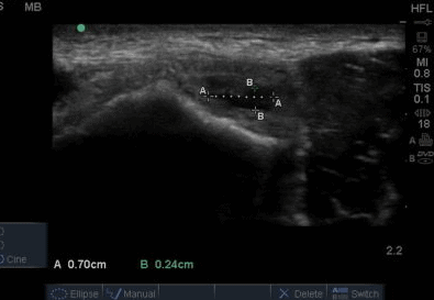
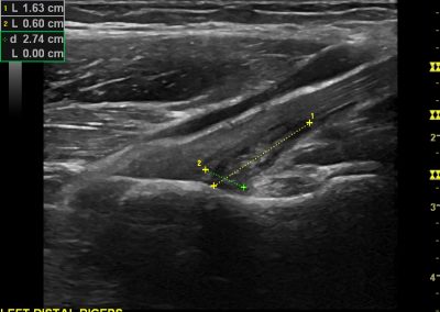
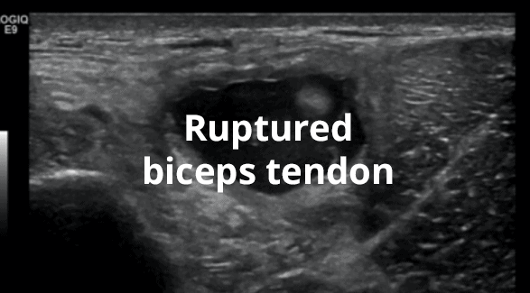
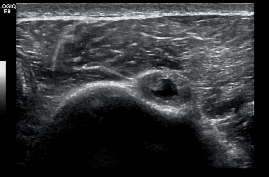
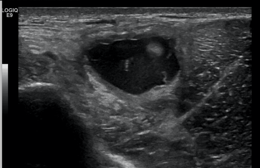
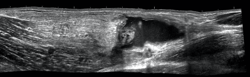
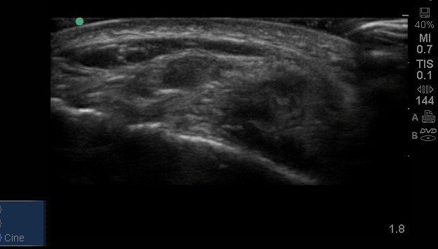
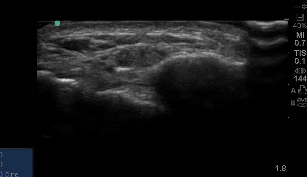
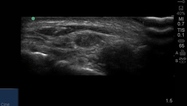
While the elbow may appear to be very accessible, it is a difficult joint to scan accurately, not least because much of the anatomy travels in planes that make good visualisation of structures challenging. In particular, the distal biceps tendon can be notorious difficult to gain good visualisation.
As with all anatomical regions, a sound knowledge of the local anatomy and anatomical relationships is essential.
Case Studies
We have developed a handful of case studies to hopefully illustrate the use of ultrasound in a clinical setting.
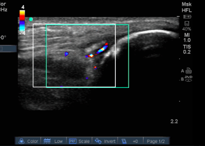
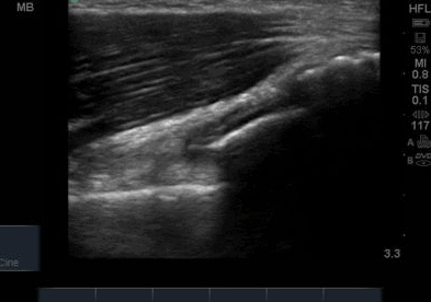
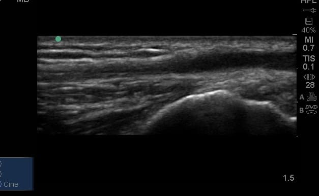
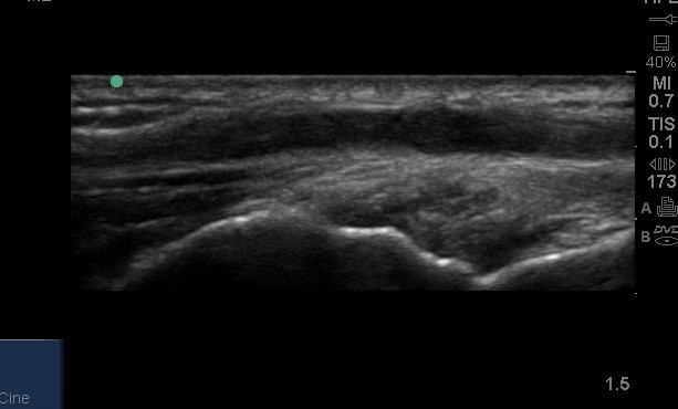
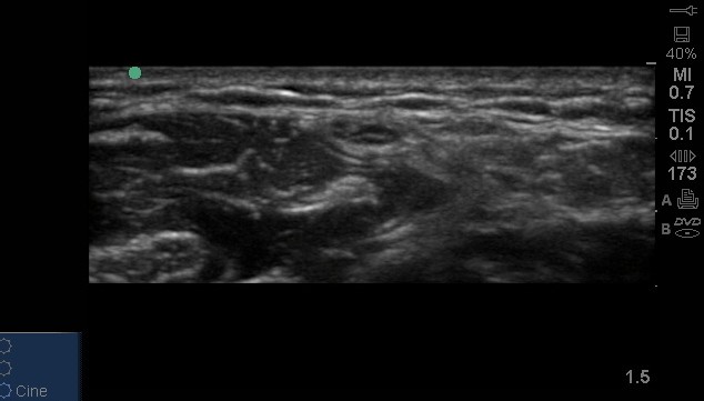
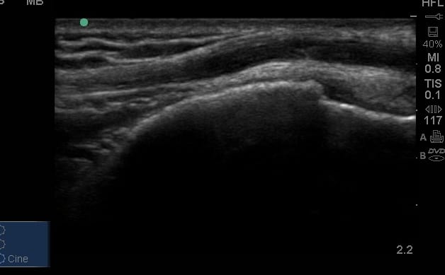
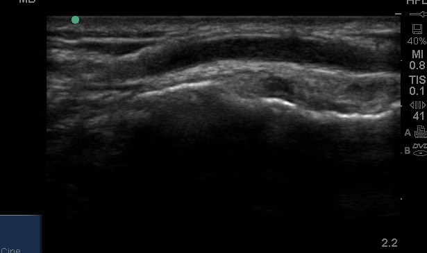
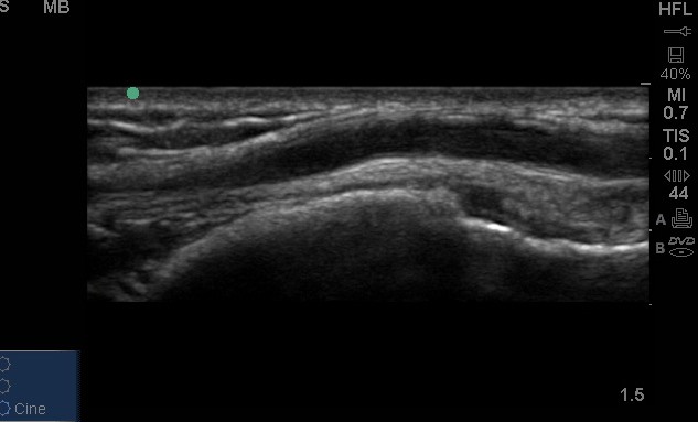
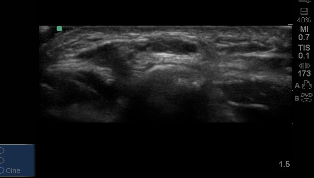
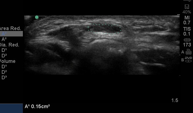
Gallery

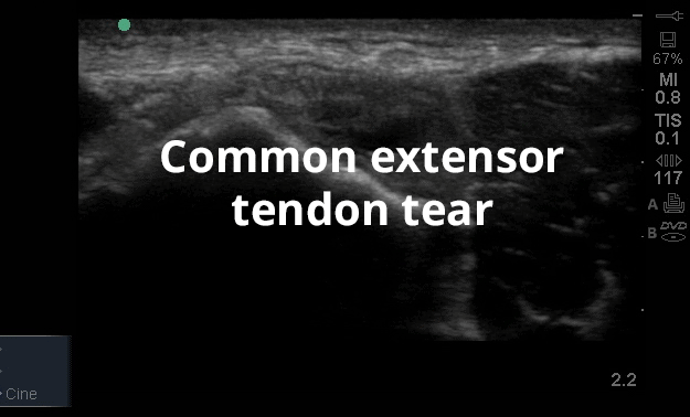

Ultrasound pathology videos
Free access MSK ultrasound articles
Register to receive our newsletter including case studies
Anatomy
The elbow joint is comprised of three articulations.
The radio capitellar, proximal radioulnar and trochlear ulnar.
These joints share a common joint capsule. The joints are supported by a number of soft tissue structures that are outlined in the dissection video. These include the medial collateral ligament,lateral collateral ligament and then the muscles and tendons of the anterior, medial, lateral and posterior aspects of the joint.