Hip ultrasound
What structures can we see at the hip?
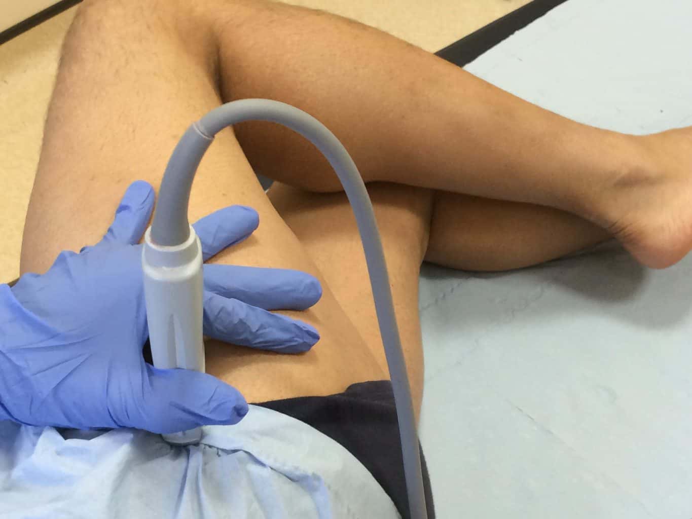
Having a good understanding on how to optimise your image through alterations in focal zone, frequency and gain is important for this region. Utilising a curvilinear for joint visualisation is often required, if available. Common regions to interrogate are the lateral hip structures including the gluteal tendons and bursae, and also the anterior recess of the joint to evaluate for effusions.
Anatomy
There are a number of ligamentous structures surrounding the joint, providing further stability. Extracapsular ligaments include Iliofemoral, ischiofemoral, and pubofemoral ligaments. Intracapsular ligaments include ligamentum teres. The hip is supplied by blood from the medial circumflex femoral and lateral circumflex femoral arteries.
There are several muscles anteriorly and these are often divided into deep – rectus femoris and Iliopsoas and pectineus, and superficial – sartorius and tensor fascia latae. Medially the adductor muscles dominate, along with the gracilis which is the most medial. Laterally, in the region of the greater trochanter, where pain is a common and a challenging clinical presentation, we find the gluteus medius, minimus and maximums, and the iliotibial tract. The gluteus maximus dominates the posterior aspect of the hip.
The femoral triangle is an important anatomical location, formed by the adductor longus medially, and the medial edge of sartorius laterally. It contains the common femoral artery and vein, the femoral nerve and inguinal lymph nodes.
We hope you find the video helpful and please do look at the product review section to see what anatomy text books we recommend. Anatomy really does provide a core foundation for using MSK ultrasound.