Ultrasound Case Study
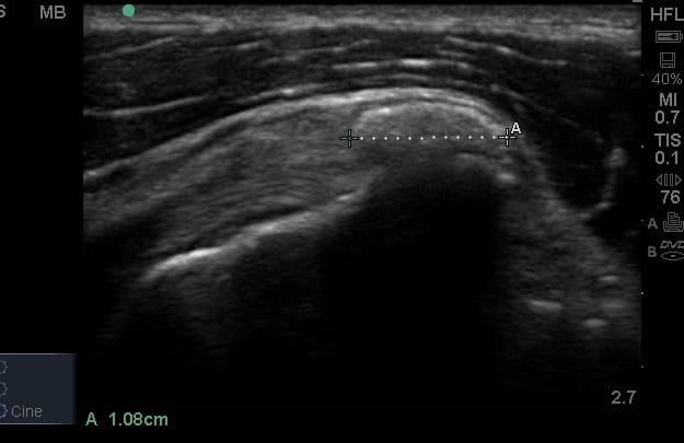
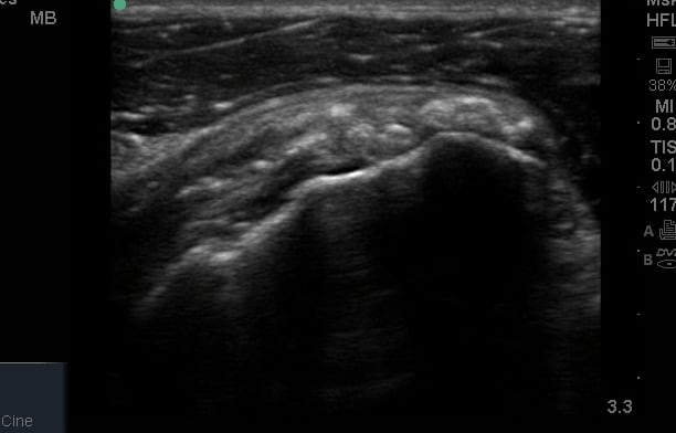
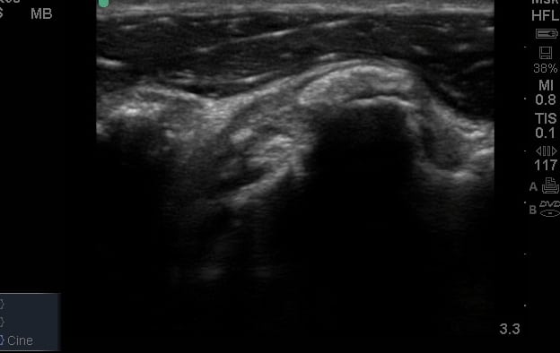
Subscapularis tendon calcification
This patient presented with a 2 year history of chronic right anterior shoulder pain, easily reproducible on impingement tests and horizontal adduction. There had been no response to Physiotherapy or a blind subacromial subdeltoid bursa corticosteroid injection.
Ultrasound demonstrated , a dense , hyperechoic structure on the distal insertion of the Subscapularis to the lesser tuberosity. This appeared to cause pain as it was approximated with the coracoid process during horizontal adduction. There is a video of this, in the video section on the shoulder page.
When visualising calcification on ultrasound, it is important to be critical of the relevance. It is widely accepted that calcific tendinopathy is present in asymptomatic shoulders. The use of dynamic sonographic evaluation can be very helpful to correlate the movement of different tissues to symptom response. There are still many question surrounding the relevance of calcific findings in ultrasound. Some useful references are shared below, and there is also an inforgraphic which is free to download on the shoulder page.


0 Comments