Ultrasound Case Study
Partial thickness rotator cuff tear: A summary
Dave Baker, Extended Scope Physiotherapist/MSK Sonographer (@davebakerphysio)
This article provides a short summary of partial thickness rotator cuff tears, this is not exhaustive and aims to be a condensed summary for reference.
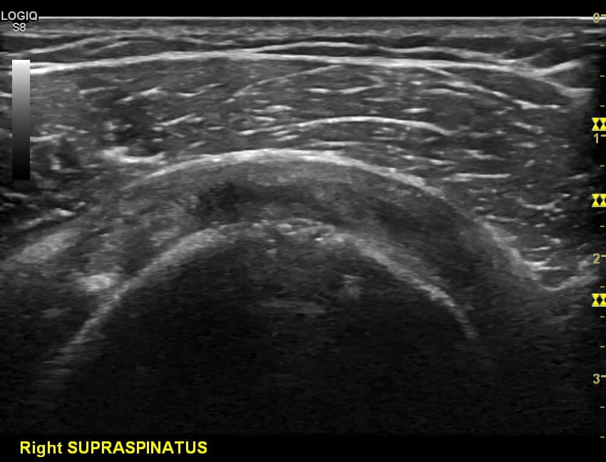
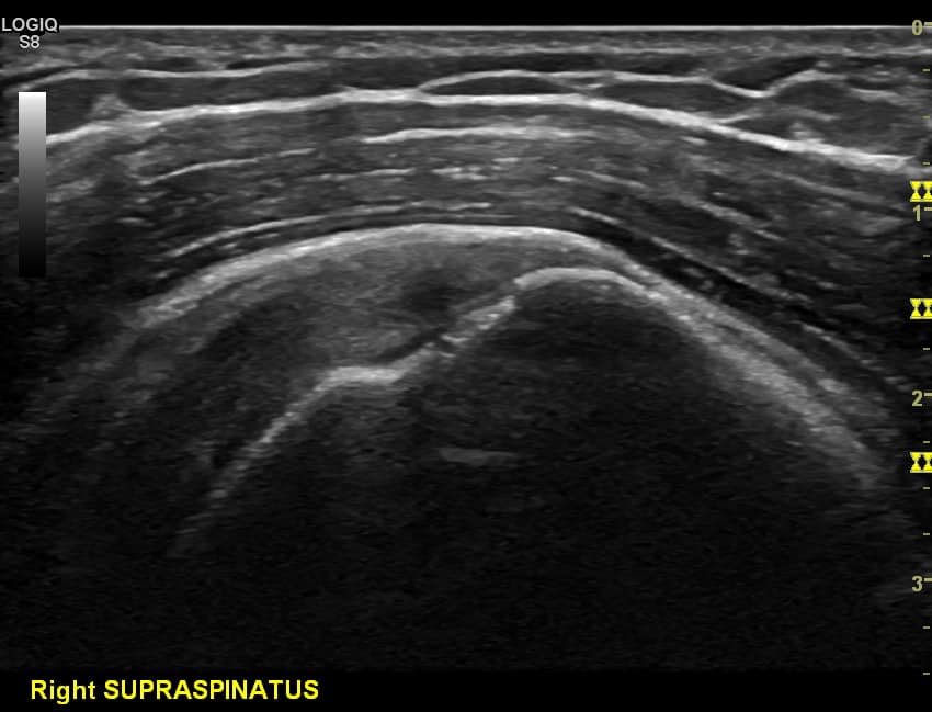
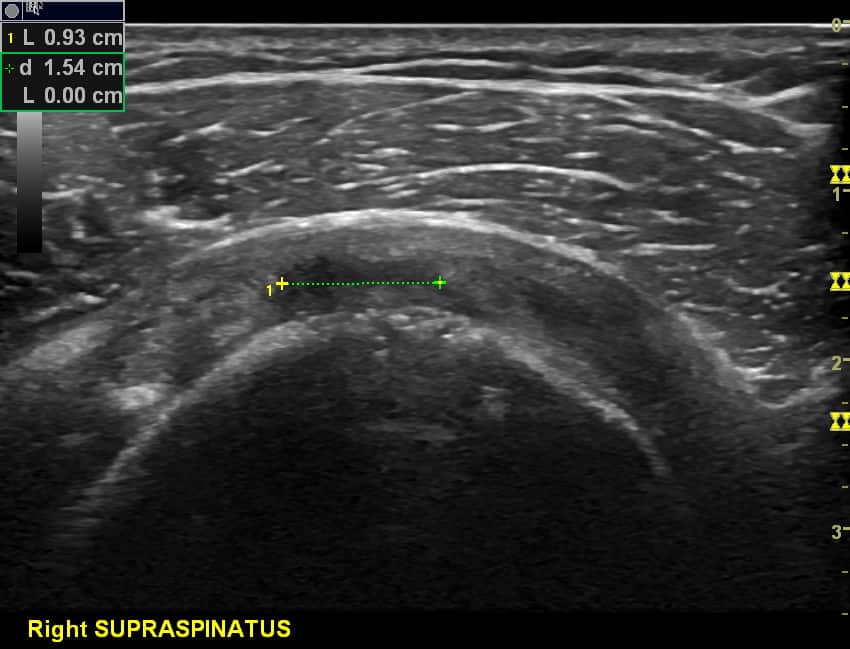
The rotator cuff (RC) comprises of four muscles which all arise from scapular and insert onto the humerus. Of these, the supraspinatus is the most common, accounting for approximately 62% of all tears (Bianchi 2007) due to local anatomy, poor blood supply and mechanical stresses related to movements of the shoulder. A key distinction when considering rotator cuff pathology is between full thickness tear, whereby the tear is through the entire tendon (Figure 1) or partial thickness rotator cuff tear (Figure 4), whereby only one aspect or surface of the tendon tears without penetration through the entire tendon thickness (Jacobson 2008).
PTRCT account for approximately 13-18% of all rotator cuff tears and are more common in younger patient group than full thickness tears. A systematic review of cadaver studies suggest the incidence of PTRCTs to be approximately 18.5% (mean age 70 years) (Reilly et al 2006) with around 30-50% being asymptomatic. Different rotator cuff tear characteristics suggest different mechanisms of lesion e.g. tears relating to chronic attrition usually present at lateral fibre insertion whereas acute tears in younger normally in younger population occur more proximal within the tendon (Jacobson 2008, Bianchi 2007).
Terminology and Classification
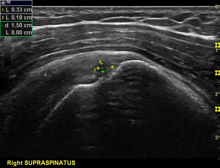
PTRCT’s are generally described by their position within the tendon i.e. for supraspinatus tendon tears on bursal (superficial) or articular (deep) surface or intra-tendinous (within the tendon not communicating with either surface (Figure 3). Articular surface tears (Figure 4) are approximately 3:1 more common due to complex composition of tendon, ligament and capsule versus tendon only composition of bursal surface. The deep fibres are less plastic and thus have less elongation potential (Vlychou et al 2009, Bianchi 2007).A common focal area for supraspinatus tears is on the articular surface at the far distal insertion of fibres immediately adjacent to the greater tuberosity of the humerus (Jacobson et al 2004, Bianchi 2007) . These are known as ‘Rim rent tears’.
Other common articular surface PTRCT include ‘PASTA’ lesions (partial articular supraspinatus tendon avulsion (Milstein and Snyder, 2003)and ‘ASFL’ (articular sided footprint lesions). Ellman (1990) graded PTRCTs into grade I (< 3mm), grade II (3mm to 6mm) and grade III (>6mm) and Ellman and Gartsman (1993) by shape.
1 – Crescent
2 – Reverse L
3 – L shaped
4 – Trapezoidal
5 – Massive tear Full thickness rotator cuff tears
Habermeyer et al (2008) proposed a two-dimensional classification for articular surface tears system grading and measuring longitudinal and sagittal extensions with reference to key anatomical structures to aid clinical decision making such as referral for surgery.
Although there is little clear guidance on overall management of partial thickness tears there is some consensus that tears involving 50-70% of the tendon width will require surgical intervention (Kamath et al 2009).
References
- Bianchi, S. Marinoli, C., (2007) Medical Radiology, Diagnostic Imaging : Ultrasound of the Musculoskeletal System. Springer. ISBN 978-3-540-42267-9
- Ellman, H., (1990)Diagnosis and treatment of incomplete rotator cuff tears. Clin Orthop Relat Res, 254, pp. 64-74.
- European Federation of Societies for Ultrasound in Medicine and Biology – Clinical Safety Statement for Diagnostic Ultrasound (2011)
- Jacobson, J.A., (2008) Musculoskeletal Ultrasound. Saunders. Elsevier Limited. ISBN 10-1-4160-3593-1
- Kamath, G., Galatz, L.M., Keener, J.D., Teefey, S., Middleton, W., Yamaguchi, K., (2009) Tendon Integrity and Functional Outcome After Arthroscopic Repair of High-Grade Partial-Thickness Supraspinatus Tears. The Journal of Bone and Joint Surgery. May01, Vol 91, Issue 5.
- McGee, D., (2007) Orthopaedic Physical Assessment 5th Edition. Saunders. ISBN-10: 0721605710
- Millstein, E., Snyder, S., (2003) Arthroscopic management of partial , full thickness, and complex rotator cuff tears : indications , techniques and complications, Arthroscopy, 19, pp.189-199
- Mitchell, C., Adebajo, A., Hay, E., Carr, A., (2005) Shoulder Pain : Diagnosis and Management in Primary Care. British Medical Journal (BMJ) November 12; 331 (7525) 1124-1128.
- Reilly, P., Macleod, L., Macfarlane, R., Windley, J., (2006) Dead men and radiologists don’t lie: a review of cadaveric and radiological studies of rotator cuff prevalence. Annals of Royal College of Surgeons England, 88, 116-121
- Rutten, M., Maresch, B,J., Jager, G.J., Blickman, J.G., van Holsbeeck, M.T., (2007) Ultrasound of the Rotator Cuff with MRI and Anatomic correlation. European Journal of Radiology Vol 62, Issue 3, pp427-436
- Rutten, M., Spaargaren, G., van Loon, T., de Waal Malefijit, L., Kiemeney, M., Jager, G., (2010) European Radiology, 20, pp. 450-457
- Teefey, S.A., Middleton, W.D., Payne, W.T., Yamaguchi, K., (2004) Detection and Measurement of Rotator Cuff Tears with Sonography; Analysis of Diagnostic Errors
- Teefey, S.A., Middleton, W.D., Yamaguchi, K., (1999) Shoulder Sonography. State of the Art. Radiol Clin North Am. 37 (4); 767-785,ix
- Teefey, S.A., Rubin, D.A., Middleton, W.D., Hildebolt, C.F., Leibold, R.A., Yamaguchi, K., (2004) Detection and Quantification of Rotator Cuff Tears. The Journal of Bone and Joint Surgery (American) 86
- Van Holsbeeck, M.T. Introcaso, J.H., (2001) Musculoskeletal Ultrasound 2nd Edition. Mosby. ISBN-10: 0323000185
- Vlychou, M., Dailiana, Z., Fotiadou, A., Papanagiotou, M., Fezoulidis, V., Malizos, K.N., Symptomatic Partial Rotator Cuff Tears; Diagnostic Performance of Ultrasound and Magnetic Resonance Imaging with Surgical Correlation. Acta Radiologica; 1; 101-105.
We hope you have found some useful learning points from this article, please continue to share with colleagues.
All feedback welcome at [email protected]


0 Comments