About this course
At The Ultrasound Site, we are passionate about the need for clinical reasoning alongside ultrasound imaging – eg what does it mean to the patient journey?. As well as our other courses on skill acquisition of learning how to perform an ultrasound scan, we wanted to develop a course which tackles some of the arguably harder questions of appropriately integrating this unique tool into clinical practice.
This one day course is aimed at musculoskeletal clinicians who have a keen interest in the use of diagnostic ultrasound for shoulder pathology. This may be useful for those wishing to gain an understanding of what ultrasound can visualise and the clinical relevance. The course aims to facilitate the integration of ultrasound imaging into your clinical practice covering practicals of shoulder ultrasound techniques, discussion of the evidence around particular pathologies and an appraisal of the evidence behind guided injection techniques in this region. There will also be the opportunity to learn approaches for ultrasound guided procedures, facilitated by The Ultrasound Site teaching faculty.
A strong clinical focus will be maintained throughout the day. The morning will firstly include a session with short presentations from experienced faculty members on calcific tendinopathy, and rotator cuff and bursal pathology. This will be followed by a practical session covering basic and more advanced shoulder ultrasound protocols.
In the afternoon, two further presentations on bony changes of the glenohumeral joint and ‘The Shoulder: To inject or not to inject?’ from Jeremy. Finally, there will be a practical session offering delegates the opportunity to learn how to approach ultrasound guided shoulder injection techniques. This will provide a useful learning experience for those considering developing ultrasound guided injection approaches or currently performing landmark guided procedures.
Who is this course suitable for?
This course is appropriate for clinicians performing diagnostic ultrasound scans and injections and also those who refer for them.
Course outline
| Time | Activity |
|---|---|
| 8.45am | Meet and registration |
| 9am | Introductions |
| 9.30am | Calcification of the rotator cuff - presentation and open forum discussion -Stuart Wildman |
| 10.15am | Rotator cuff and bursal changes - presentation and open discussion - with Dave Baker |
| 11.00 | Break |
| 11.15am | Demonstration and practicals in small groups - ultrasound evaluation of structures around the shoulder girdle. Including small groups and direct tutor feedback |
| 12.45 | Lunch |
| 1.15/1.30pm | The role of ultrasound in evaluating bony pathology at the shoulder - presentation and open forum discussion - with Robert Mast |
| 2.15pm | The Shoulder: To inject or not to inject?presentation and open forum discussion - Dr Jeremy Lewis |
| 3pm | Break |
| 3.15pm | Practical and demonstration of approaches to ultrasound guided shoulder injections. Including small groups and direct tutor feedback |
| 4.30 | Summary and discussion for the day |
| 4.45/5 | Finish |
Course Dates & Booking
If your attendance on the course is to be funded by your trust or you have any other queries then please contact us.
| Date | Places | Price | |
|---|---|---|---|
| Date | Places | Price |

Become a free member and receive access to our exclusive content & updates
Your Course Tutors
Mr Stuart Wildman
MSc, BSc (Hons) Physiotherapy, HCPC, MCSP, PG Cert/CASE accredited Musculoskeletal Sonography, PG Dip injection therapy
Stuart is an Extended Scope Physiotherapist and MSK Sonographer working in the NHS in London. He has an interesting working week, dividing his time between Radiology and Physiotherapy, here he performs both diagnostic and guided interventions in both. He qualified from the University of Southampton in 2003 with a BSc Physiotherapy, and went on to gain an MSc in Advanced Neuromusculoskeletal Physiotherapy at The University of Hertfordshire and a PG Cert in MSK Sonography at Canterbury and Christ Church University.
Mr Robert Mast
BSc Physiotherapy HCPC, MCSP, PgCert Musculoskeletal Sonography, Non Medical prescriber
Rob trained and qualified as a physiotherapist through the HvA in Amsterdam in 1993.
He has been working for Homerton University Hospital as an Extended Scope Physiotherapist in the primary care setting since 2004. He works one day per week in the Radiology Department of Homerton University Hospital as an MSK sonographer both for diagnostics as well as for carrying out interventional procedures.
Mr David Baker
BSc Physiotherapy HCPC, MCSP, PG Cert Musculoskeletal Sonography
David brings together his unique skills as ESP, MSK Sonographer, experienced injection therapist, and lecturer to the website. David works as an Extended Scope Physiotherapist in the NHS, working extensively with diagnostic ultrasound since 2007 and studied for his formal Pg Cert at Canterbury Christ Church University. He has further developed his special interest in ultrasound guided injection therapy.
![]()
Complete this course to access The Ultrasound Site private course group on MedShr
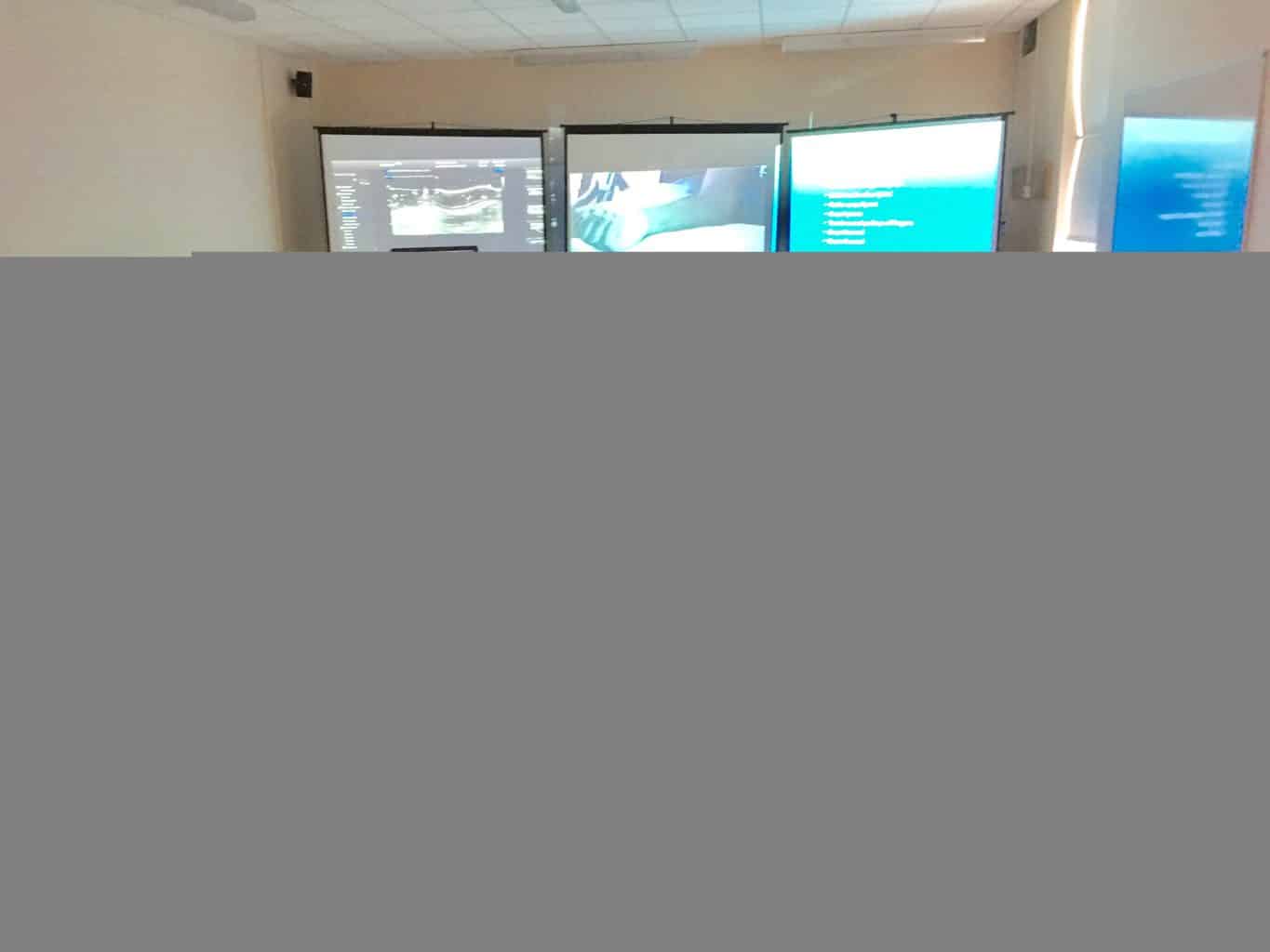
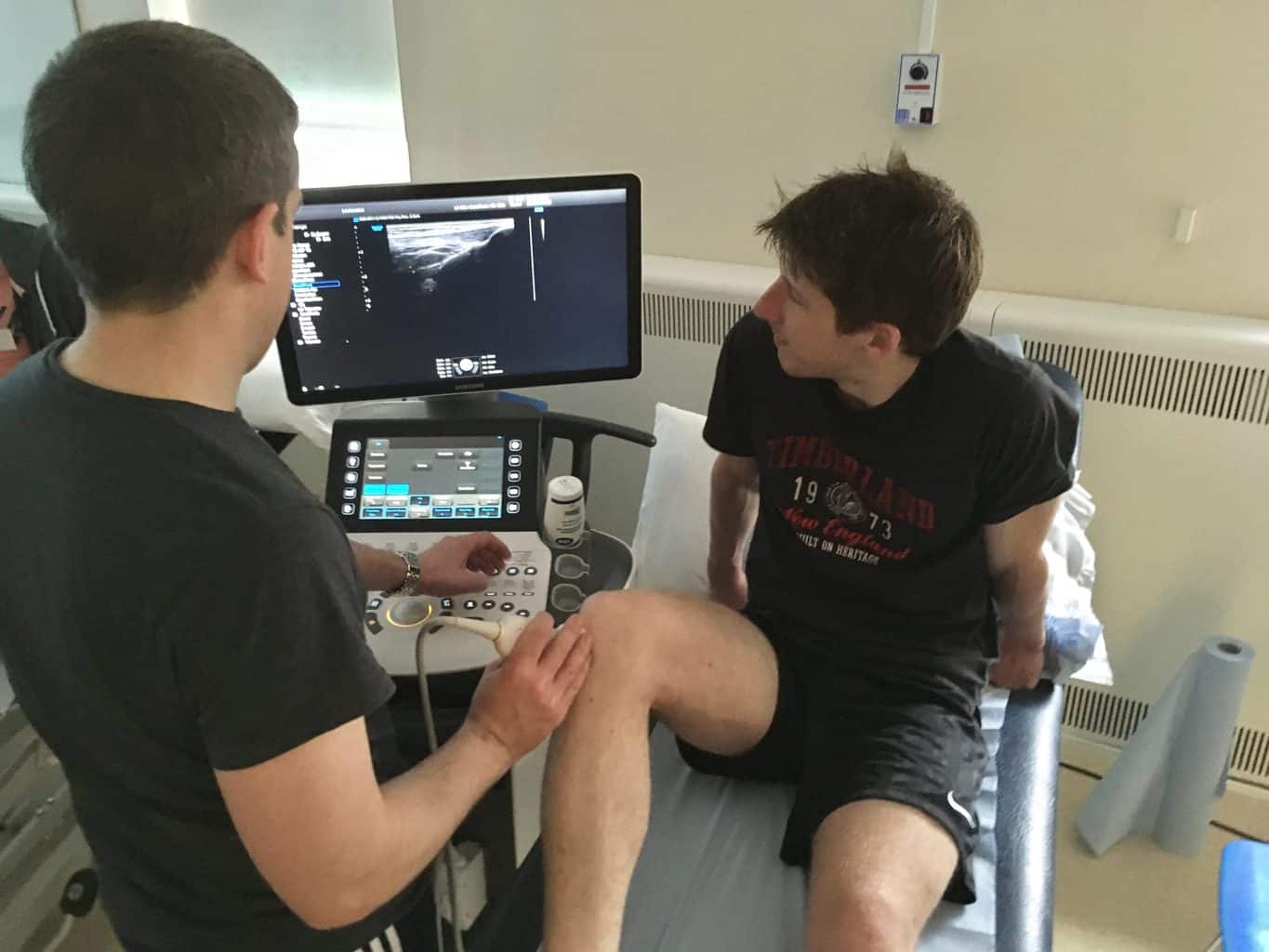
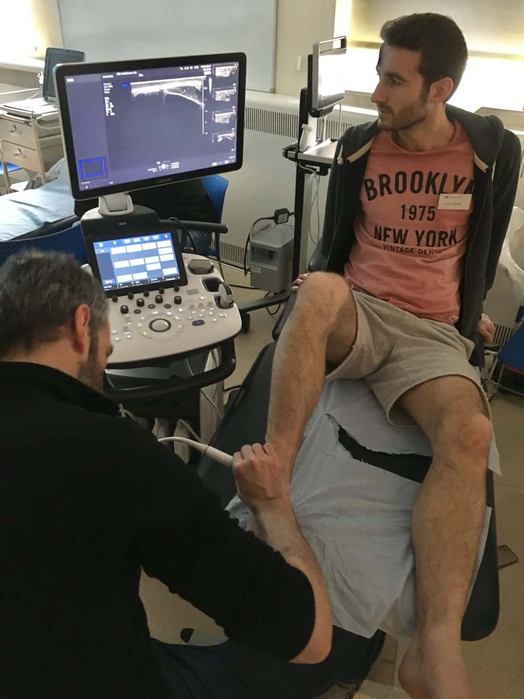
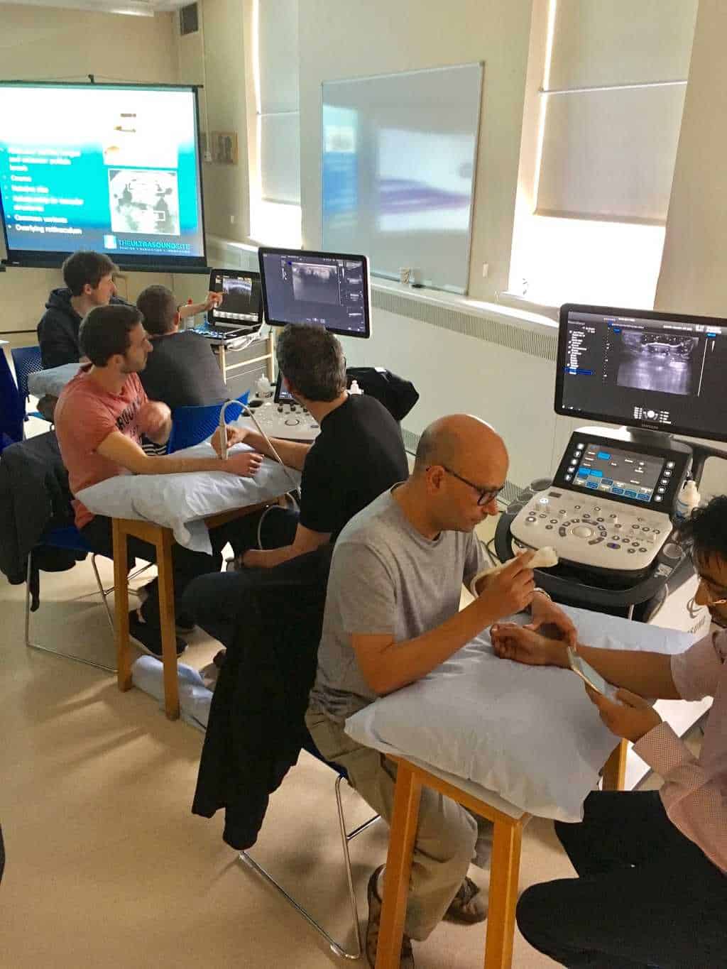
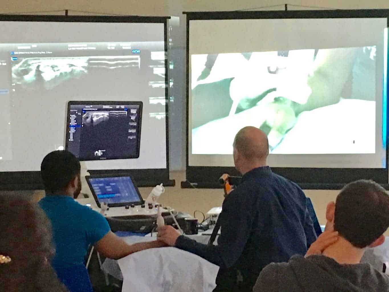
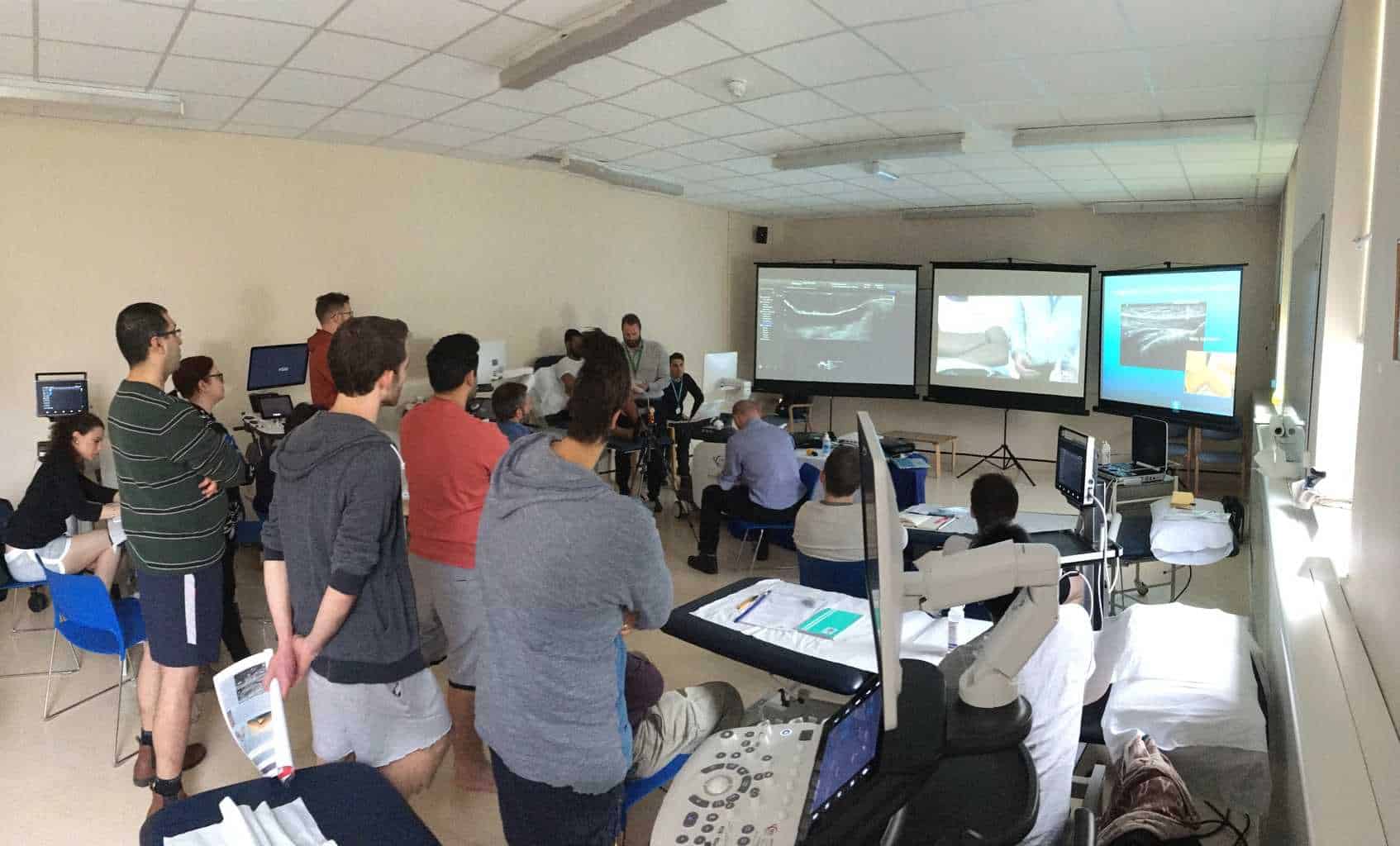
Previous Course Photo Gallery
FAQs
Q. How do I know that this course is of high quality?
All the lecturers on the course have completed/training towards a PG Cert in MSK Sonography and have been using MSK Ultrasound for a substantial amount of time. They are also Extended Scope Physiotherapists with many years experience of integrating ultrasound into their practice alongside other forms of imaging such as MRI. Several teach on PG Cert MSK Ultrasound courses as guest lecturers. The lecturers are also members of executive committees of CSP Professional Networks so are very familiar with MSK ultrasound development within Physiotherapy, and the use of Injection Therapy.
Q. Do we get to use the ultrasound machines?
We know the importance of hands-on practice when scanning! So we can reply with a massive YES! The course is designed with high student-to-lecturer ratios and large number of machines to ensure that as much time as possible is spent with the students scanning under close supervision. We understand the subtlety of moving a probe to ensure you achieve a high quality diagnostic image.We also use crib sheets, which are signed off at the end of each body part to ensure that everybody has an opportunity to scan each of the listed areas under each body part.
Q. Does this qualify me as a sonographer?
Surprisingly, a sonographer is not a protected title and therefore anyone can theoretically call themselves a ‘sonographer’. I guess it is more a question of does this deem me competent to perform ultrasound in the setting I wish to use it in?. Well, this depends on the setting and the local governance but essentially this course is designed as an important first step for people who are considering an ultrasound qualification, or who are interested in learning ultrasound to give them an idea of how ultrasound may fit into their practice. The course can also serve as a refresher, perhaps for certain joints.
To undertake a formal qualification which would lead you to be able to work in for example Radiology departments within the NHS you would need to progress on to complete a PG Cert qualification on a CASE accredited course. This course would place you in a strong position before starting your CASE course.
Q. How do I know if I am scanning things correctly?
Because we have a large ratio of tutors to students, there will be tutors with each group for as much time as possible throughout the day to ensure that you are handling the probe correctly and there are lots of tips on image optimising and use of the machine technology to get the best image! Basically, we will be peering over your shoulder! We also use a competencies based crib sheet to ensure that everybody has scanned all of the importance structures on each body part area.
Q. What are the advantages of doing this course?
The advantages of doing this course is that all of the tutors have extensive experience of working with ultrasound and ultrasound guided injections working within a busy multidisciplinary team physiotherapy outpatient department.
Q. What are the terms and conditions for refunds and bookings?
All details for the refund policy and bookings terms and conditions can be found HERE
Q. Do I need to have scanned before in order to attend a course?
No. We have many people who attend the course who have little, or no, previous scanning experience. This course is designed to accommodate people who have no previous scanning experience and are interested in learning from scratch. However, as mentioned above, it is also a very useful learning experience for people with some scanning background as well.
Q. Do you provide training to whole departments?
We are able to travel and provide a customised training program for a group of clinicians in a department. For further discussion email us at [email protected]
Q. Does this course qualify me to perform ultrasound guided injections in my workplace?
No. In order to perform image guided injections in your workplace you must conform to local guidelines and competency requirements. This course, however, may form part of your CPD and learning if you are looking to start performing ultrasound guided injections, or as a top-up course as part of your continuing CPD.
Course location
Human Anatomy Unit ,Rm 14L10, 14th Floor, Laboratory Block , Charing Cross Hospital Campus ,W6 8RP