Introduction to diagnostic MSK ultrasound course
Course Dates & Booking
All prices below are inclusive of VAT
If your attendance on the course is to be funded by your trust or you have any other queries then please contact us.
| Date | Places | Price | |
|---|---|---|---|
| 21st November 2026 - Introductory MSK ultrasound course - Day 1 (Lower Limb) | 15 in stock | £340.00 | |
| 22nd November 2026 - Introductory MSK ultrasound course - Day 2 (Upper limb) | 14 in stock | £340.00 | |
| 21/22 November 2026 - Introduction to MSK ultrasound course | 12 in stock | £599.00 | |
| 27th February 2027 - Introductory MSK ultrasound course - Day 1 (Lower Limb) | 15 in stock | £340.00 | |
| 28th February 2027 - Introductory MSK ultrasound course - Day 2 (Upper limb) | 15 in stock | £340.00 | |
| 27/28 February 2027 - Introduction to MSK ultrasound course | 14 in stock | £599.00 | |
| 12th June 2027 - Introductory MSK ultrasound course - Day 1 (Lower Limb) | 15 in stock | £340.00 | |
| 13th June 2027 - Introductory MSK ultrasound course - Day 2 (Upper limb) | 15 in stock | £340.00 | |
| 12/13 June 2027 - Introduction to MSK ultrasound course | 14 in stock | £599.00 | |
| 20th November 2027 - Introductory MSK ultrasound course - Day 1 (Lower Limb) | 15 in stock | £340.00 | |
| 21st November 2027 - Introductory MSK ultrasound course - Day 2 (Upper limb) | 15 in stock | £340.00 | |
| 20/21 November 2027 - Introduction to MSK ultrasound course | 14 in stock | £599.00 | |
| Date | Places | Price |
About this course
Join our highly regarded Introduction to diagnostic MSK Ultrasound course, a CPD course trusted by hundreds of MSK clinicians from all over the globe for over a decade. As one of the most established courses available, we have provided the essential introduction to this topic for countless professionals, helping to shape clinical practice across the UK and beyond. This highly practical two-day musculoskeletal ultrasound course is your first step to becoming a competent and confident MSK sonographer.
Our MSK ultrasound course is led by highly experienced, full-time musculoskeletal clinicians, including Advanced Practice Physiotherapists and Consultant MSK Sonographers. With over a decade of experience using musculoskeletal ultrasound in busy NHS and private outpatient MSK departments, as well as in Radiology MSK clinics, our tutors provide unparalleled, real-world insights.
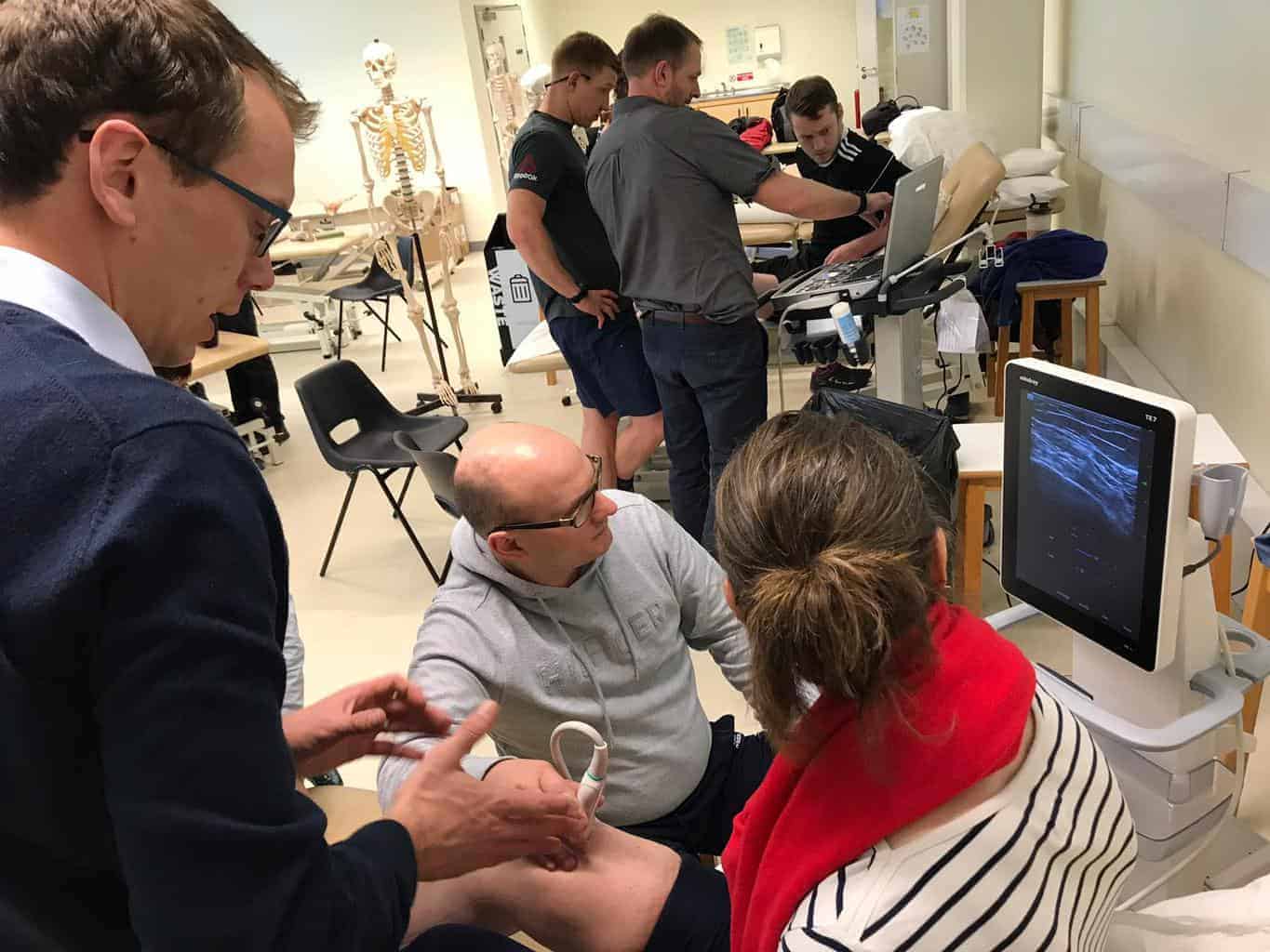
This introductory MSK ultrasound course is ideal for beginners and has been the starting point for hundreds of clinicians over the last 10 years. Our proven formula for success prioritises hands-on learning, with a high tutor and scanner to student ratio (maximum 3 per system) to ensure you get ample scanning time and close support. The course covers the scanning of all peripheral joints and includes short lectures on essential topics such as basic physics, pathology case studies, and integrating ultrasound into your clinical practice. We also provide an overview of clinical governance and regulation, guidance on presenting a business case for ultrasound, an introduction to ultrasound-guided injections, and a critical appraisal of ultrasound as a diagnostic tool.
To further enhance your learning, several lectures are available 24/7 online to complement your face-to-face training.
Discover why hundreds of clinicians have chosen our highly regarded Introduction to diagnostic MSK ultrasound course to transform their practice.
This comprehensive musculoskeletal ultrasound course takes place at the Mary Seacole Building, Brunel University London, 8 Kingston Ln, Uxbridge UB8 3PN. View Location Maps
Who is the Introduction to diagnostic MSK ultrasound course suitable for?
This course is suitable for allied health or medical professionals who:
- Have experience of working with patients with musculoskeletal injuries or complaints
- You must be able to use diagnostic MSK ultrasound within your professional scope of practice and insured to do so.
- Relevant medical or allied health professional experience or qualifications.
- Current registration with the relevant UK professional body eg HCPC or GMC, or foreign equivalent if travelling from abroad
Who is this course endorsed by?
European Society of Musculoskeletal Radiology for 1 point Category 2
The Royal College of Radiologists (RCR) This course provides 14 CPD credits in accordance with the CPD Scheme of the Royal College of Radiologists.
Planning Your Visit: Travel & Accommodation
Planning your journey to our course at Brunel University, Uxbridge (UB8 3PN) is straightforward, thanks to its excellent transport links. For our attendees from across the globe, the campus is conveniently located just a 15-minute taxi or bus ride from London Heathrow Airport (LHR). The university is also easily accessible via public transport; Uxbridge Underground Station is the terminus for both the Metropolitan and Piccadilly lines, providing direct access from central London and major rail hubs like King's Cross St Pancras. For those driving, the campus is situated near the M25, M40, and A40 motorways. There is parking available on-site, a short walk from the venue.
You will find a wide range of accommodation options nearby. Please note: The following hotels are listed as helpful suggestions only. We do not have a formal partnership with them and take no responsibility for your booking or the quality of your stay.
For maximum convenience, the Lancaster Hotel & Conference Centre is located directly on the Brunel campus. Nearby in Hillingdon, The Red Lion Hotel offers a more traditional setting. Additionally, Uxbridge town centre, a short bus, walk or taxi ride away, has several reliable chains, including a Premier Inn and a Travelodge.
We recommend booking your accommodation well in advance to secure the best rates, ensuring a comfortable and stress-free stay while you attend our highly regarded musculoskeletal ultrasound course.
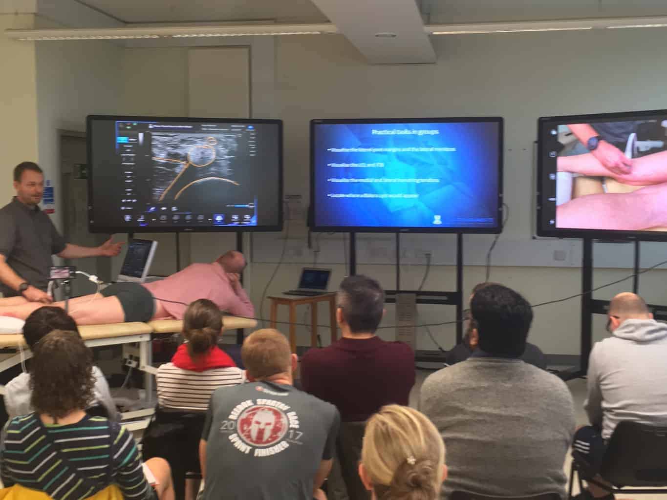
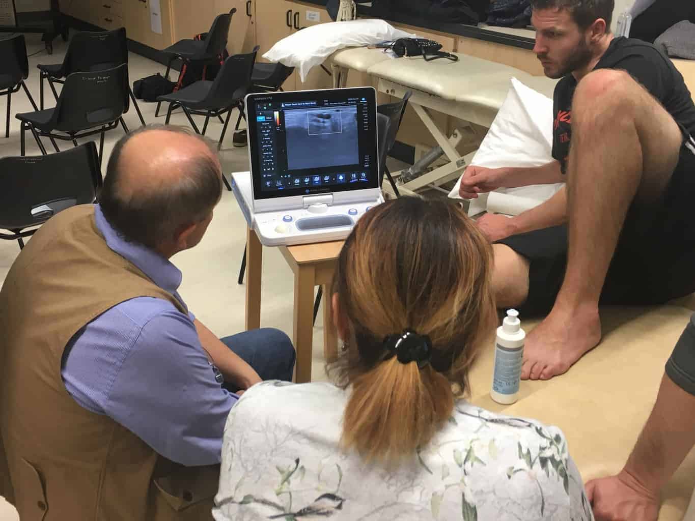
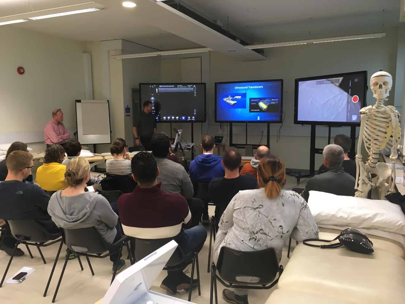
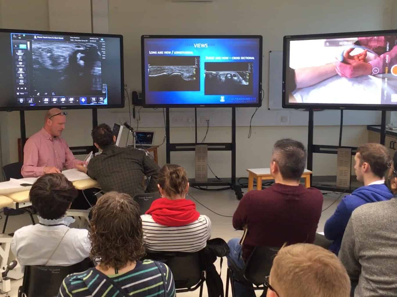
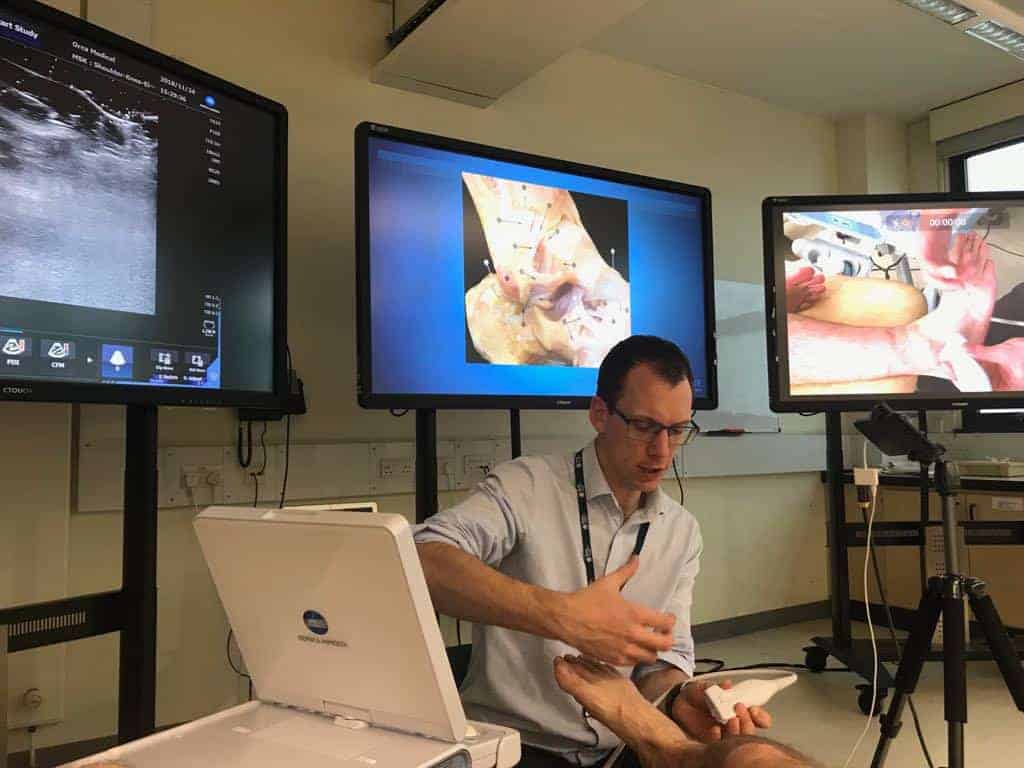
Examples of our teaching
Our CPD courses provide practical knowledge but do not award a certificate of competency (excluding the Brunel PG Cert program). You must enquire with your relevant professional body to confirm if the skills covered within our courses are considered within the scope of your professional practice, and whether your professional insurance will cover you to use them in your practice. Ultrasound regulation is a complex issue. Any skills covered by the course should be maintained and developed through ongoing mentorship, peer support, and training. Please get in touch if you have questions here .
EXTRA ONLINE LECTURES AND TUTORIAL VIDEOS
")
ONLINE MSK ULTRASOUND LECTURES 24/7
All course delegates who attend our MSK Ultrasound training courses will get unlimited access to online lectures by our course tutors. These provide a more in-depth appreciation of the topics outlined below. We are constantly adding to this selection, so keep your eyes peeled!
- An introduction to the Physics of MSK ultrasound
- An introduction to probe skills and orientation to the image
- Ultrasound system settings and knobology
- An introduction to recognising pathology on MSK ultrasound
- An introduction to quality assurance and professional issues of MSK ultrasound
GAIN ACCESS TO OUR PRIVATE FACEBOOK MSK ULTRASOUND COMMUNITY OF PRACTICE
By attending our courses you will also gain access to your tutors in our private, Facebook community of MSK Ultrasound practice with specific groups for upper and lower limb MSK Ultrasound pathology, discussing anonymised cases and articles of interest. We also have groups for upper and lower limb guided injection techniques, discuss your techniques - what went well, what could be improved and what the evidence is. Lastly, but importantly we have established groups on training and governance.
")
The next course starts in....
Day(s)
:
Hour(s)
:
Minute(s)
:
Second(s)
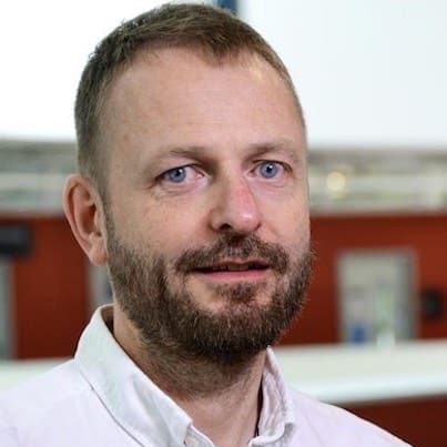
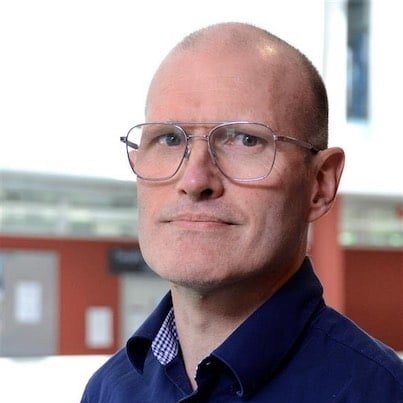
Your Course Tutors

Mr Stuart Wildman
MSc, BSc (Hons) Physiotherapy, HCPC, MCSP, PG Cert/CASE accredited Musculoskeletal Sonography, PG Dip injection therapy
Stuart is an Extended Scope Physiotherapist and Consultant MSK Sonographer working in the NHS in London. He founded The Ultrasound Site. He has an interesting working week, dividing his time between Radiology and Physiotherapy, here he performs both diagnostic and guided interventions in both. He qualified from the University of Southampton in 2003 with a BSc Physiotherapy, and went on to gain an MSc in Advanced Neuromusculoskeletal Physiotherapy at The University of Hertfordshire and a PG Cert in MSK Sonography at Canterbury and Christ Church University.
Mr David Baker
BSc Physiotherapy HCPC, MCSP, PG Cert Musculoskeletal Sonography
David brings together his unique skills as ESP, MSK Sonographer, experienced injection therapist, and lecturer to the website. David works as an Extended Scope Physiotherapist in the NHS, working extensively with diagnostic ultrasound since 2007 and studied for his formal Pg Cert at Canterbury Christ Church University. He has further developed his special interest in ultrasound guided injection therapy.
Mr Robert Mast
BSc Physiotherapy HCPC, MCSP, PgCert Musculoskeletal Sonography, Non Medical prescriber
Rob trained and qualified as a physiotherapist through the HvA in Amsterdam in 1993.
He has been working for Homerton University Hospital as an Extended Scope Physiotherapist in the primary care setting since 2004. He works one day per week in the Radiology Department of Homerton University Hospital as an MSK sonographer both for diagnostics as well as for carrying out interventional procedures.
Introduction to diagnostic MSK ultrasound course outline
Day 1 (Lower Limb)
| Time | Session | Activity |
|---|---|---|
| 08:45 - 9.00 | Registration, introductions and coffee. | |
| 9.00-9.30 | Basic Physics, Acknowledging artefacts, image optimisation. Safe use of ultrasound - inc thermal/non-thermal effects | Presentation |
| 9.30-10.30 | Image optimisation practical, probe skills and identifying structures. | Practical session and demonstration - small groups, high specification US system. |
| 10.30 | Break - Coffee and biscuits! | |
| 10.45 -12.30 | Knee – scanning techniques, demonstration, facilitated group working, examples of common pathology. Structures visualised include the suprapatellar recess, extensor mechanism, following the quads from proximal to distal, patella tendon, MCL, medial meniscus, pes anserine complex. LCL, lateral meniscus, ITB insertion. Hamstrings and their insertions. | Practical session and demonstration - small groups, high specification US system. |
| 12:30-13:15 | Lunch | |
| 13-15 - 15.00 | Ankle and Foot – scanning techniques, demonstration, facilitated group working and examples of common pathologies. Structures visualised include Achilles tendon, Tibialis Posterior, Flexor Hallucis, Tibialis Anterior, Extensor digitorum,Anterior recess of the talocrural joint. lateral ankle ligaments. | Practical session and demonstration - small groups, high specification US system. |
| 15.00 | Break - Coffee and biscuits! | |
| 15.15-16.15 | Hip – scanning techniques, demonstration, facilitated group working, examples of common pathology, and techniques. Structures include anterior hip recess, gluteal tendons and insertions, trochateric bursa. | Practical session and demonstration - small groups, high specification US system. |
| 16.30 | Finish |
Day 2 (Upper Limb)
| Time | Activity |
|---|---|
| 09:00 - 09:30 | Discussion on preparing a business case, regulation of ultrasound use and quality assurance. |
| 9:30 - 10:15 | Revision of previous days techniques |
| 10:15-10:30 | Break |
| 10.30 – 12.30 | Shoulder – scanning techniques, demonstration, facilitated group working, examples of common pathology. Visualisation of Long head of biceps and the sheath, Subscapularis, Supraspinatus, Infraspinatus, Subacromial bursa, the posterior recess of the joint, AC joint |
| 12.30 -13.15 | Lunch |
| 13.15 – 15.00 | Wrist and Hand – scanning techniques, demonstration, facilitated group working, examples of common pathology. Visualisation of the key tendinous structures of the volar aspect of the wrist, alongwith the median nerve and other neural and vascular structures. The six extensor compartment of the dorsum of the wrist. Finger anatomy and discussion of common pathology including trigger finger and OA changes. |
| 15:00-15:15 | Break |
| 15.15 -16.15 | Elbow – scanning techniques, demonstration, facilitated group working, examples of common pathology. Visualisation of triceps insertion and the posterior recess of the elbow joint, common extensor tendons, common flexor tendons and insertions. The anterio aspect of the joint including distal biceps insertion, the brachial artery and median nerve. |
| 16:30 | Finish |
Previous Course Photo Gallery


Course feedback
When you come to one of our Introduction to diagnostic MSK ultrasound courses we ask for your feedback. We read this, discuss this and make changes following this. Here is a summary of data received since October 2018, and we will update this after every course to make sure it gives an accurate and honest reflection.
Overall, how would you rate the Introduction to diagnostic MSK ultrasound course?
- Excellent88.9%
- Good11.1%
- Fair50%
- Poor50%
'The Introduction to diagnostic MSK ultrasound course was informative and useful'
- Strongly agree90%
- Agree10%
- Neutral50%
- Disagree50%
- Strongly disagree50%
'The Introduction to diagnostic MSK ultrasound course met my learning objectives for attending'
- Strongly agree90%
- Agree10%
- Neutral50%
- Disagree50%
- Strongly disagree50%
'The tutors were approachable and offered feedback'
- Strongly agree100%
- Agree50%
- Neutral50%
- Disagree50%
- Strongly disagree50%
'There was a good mix of learning and practical exercises'
- Strongly agree100%
- Agree50%
- Neutral50%
- Disagree50%
- Strongly disagree50%
%
'How likely are you to attend a future course with us?'
Testimonials
"The two day introduction to MSK ultrasound is a very well planned and delivered course. Perfect blend of theory and small group practical sessions. The resources and quality of teaching are excellent. Stuart, Rob and Dave have huge amounts of experience, both as teachers and practitioners, and their enthusiasm and passion for the subject is evident. We also had the added bonus of being able to tap into Fredy’s amazing knowledge of anatomy.
The course supplies a really good grounding of knowledge in a way that can be understood what ever background profession you have. Each joint is covered firstly with anatomy and ultrasound appearance of both normal and abnormal. This is followed by a practical demonstration of scanning technique. Then the opportunity to reinforce the learning by scanning in small groups. This is all presented in a way that encourages questioning and interaction. I particularly appreciated all the practical hints and tips offered throughout the two days. I would recommend this course to anyone interested in extending their practice to include MSK ultrasound.''

"The Introductory MSK Ultrasound course was the perfect introduction to a topic that can appear daunting at first. The days are well structured with an appropriate mix of technique, anatomy, governance guidance and the physics behind the modality. Three screens are set up allowing the lecture slides, camera for probe positioning and a mirror of the ultrasound image to be viewed simultaneously.
Each section was introduced with a review of anatomy by Freddy, a Physio and anatomist, which was incredibly engaging and immediately applicable to Stuart and Rob’s demonstration of the ultrasound technique for that area. Stuart and Rob’s clinical pearls from their experience paired with their approachability made them excellent course leaders. Instructor to delegate ratio was ideal, I found that most of my scans were supervised by one of the instructors allowing invaluable personalised feedback. At no point did the course feel rushed; the pace, depth of content and practice time was perfect for an introductory level.
The introduction to MSK Ultrasound course is an excellent, accessible introduction to diagnostic ultrasound. I left feeling more confident and less intimated with an appetite for further education in this field. I would highly recommend this course to anyone seeking an introduction to diagnostic ultrasound.''

"The course was very participatory with lots of hand on experience on the ultrasound machines and a
wide content covering all the joints. The facilitators were excellent, ready to answer all questions and also provide additional instruction during the practical sessions.
I would recommend the introductory course to anyone with an interest in MSK ultrasound.."
"
'I am selective in choosing the course for my medical education .I was impressed with the solid evidence-based content, practical hands on teaching, and exclusive Problem solving tips from the instructors. '
"
"Having recently gained a place on a Post Graduate Certificate in Musculoskeletal Ultrasound (MSK US) course my objective was to explore the foundation skills of US before the start of the university course. This weekend programme is run by three highly experienced Extended Scope Physiotherapists using MSK US in an outpatient department and radiology daily.
Day one commenced with a brief but very well structured theoretical component of US. Attention was made to the importance of physics and key terminology used during US examination. The US terminology presented was new to many and therefore a clear benefit, particularly as the descriptions transcended throughout the weekend. The practical element was broken into anatomical parts. Day 1 focussed on the lower limb with emphasis on the scanning planes of the hip, knee and ankle. The rewarding aspect of the practical was the low student numbers. The course organisers make it clear the student numbers are kept low. By doing so every student can pair up thus having more time applying the US skills taught on respective partners. Each lecture provides a clear, succinct evaluation and assessment approach before ample time given for students to practice.The course leaders have clearly thought how best the practical element could be presented. With the use of undergraduate students coming to help out as models, the use of live video links to provide clear images of probe positioning and live US images also conveyed onto large screens the teaching environment is highly rewarding. There was a strong emphasis on keeping the course clinically relevant and evidence based. After each anatomical part is covered further slides presenting both common and uncommon pathological findings are discussed.
Day two commenced with a choice for students. Either have time to practice techniques taught from the previous day or listen to a lecture offered by David Baker. The one hour lecture focussed on governance issues surrounding US and offered some very useful guidance how clinicians may put together a business case to apply for their own US units in MSK services. As a clinician looking to put a case to my Trust David’s knowledge and help in this area was greatly received. The rest of the day focused on the upper limb. A significant proportion of time was rightly allocated to the shoulder as this is the most likely area US would be applied in the upper limb. The teaching approach was similar to day 1 with plenty of time to practice skills. The course was completed with a succinct evaluation of the 2 days.
I left the weekend course feeling it was undoubtingly worth the time. All 3 presenters have a clear passion for the implementation of US into MSK service provision and teaching other MSK clinicians its value. If as a clinician, you are wishing to learn the fundamentals behind the use of US in clinic or like myself looking to gain a foundation of skills to build upon before the PG Cert I would strongly encourage clinicians to look at this weekend course. Well done to Stuart, Robert and David for the time and dedication put into driving MSK US standards forward."
Course Dates & Booking
All prices below are inclusive of VAT
If your attendance on the course is to be funded by your trust or you have any other queries then please contact us.
| Date | Places | Price | |
|---|---|---|---|
| 21st November 2026 - Introductory MSK ultrasound course - Day 1 (Lower Limb) | 15 in stock | £340.00 | |
| 22nd November 2026 - Introductory MSK ultrasound course - Day 2 (Upper limb) | 14 in stock | £340.00 | |
| 21/22 November 2026 - Introduction to MSK ultrasound course | 12 in stock | £599.00 | |
| 27th February 2027 - Introductory MSK ultrasound course - Day 1 (Lower Limb) | 15 in stock | £340.00 | |
| 28th February 2027 - Introductory MSK ultrasound course - Day 2 (Upper limb) | 15 in stock | £340.00 | |
| 27/28 February 2027 - Introduction to MSK ultrasound course | 14 in stock | £599.00 | |
| 12th June 2027 - Introductory MSK ultrasound course - Day 1 (Lower Limb) | 15 in stock | £340.00 | |
| 13th June 2027 - Introductory MSK ultrasound course - Day 2 (Upper limb) | 15 in stock | £340.00 | |
| 12/13 June 2027 - Introduction to MSK ultrasound course | 14 in stock | £599.00 | |
| 20th November 2027 - Introductory MSK ultrasound course - Day 1 (Lower Limb) | 15 in stock | £340.00 | |
| 21st November 2027 - Introductory MSK ultrasound course - Day 2 (Upper limb) | 15 in stock | £340.00 | |
| 20/21 November 2027 - Introduction to MSK ultrasound course | 14 in stock | £599.00 | |
| Date | Places | Price |


Course location
FAQs
Q. How do I know that this course is of high quality?
All the lecturers on our courses have completed formal training MSK Sonography and have been using MSK Ultrasound for several years. Crucially, they are clinicians with many years experience of integrating ultrasound into their practice alongside other forms of imaging such as MRI giving a great breadth of experience to enable them to teach ultrasound in the correct clinical context.
Several teach on PG Cert MSK Ultrasound University based courses as guest lecturers, and present at different events each year. The lecturers are also members of executive committees of professional organisations such as the Chartered Society of Physiotherapy, The Royal College of Radiologists and British Association of Sports and Exercise Medicine, so are very familiar with MSK ultrasound and injection therapy development within different professional roles.
Q. Do we get to use the ultrasound machines?
We know the importance of hands-on practice when scanning! As we've been there when we were learning! So we can reply with a massive YES! The course is designed with high lecturer:student ratios, with on average a lecturer for every 5 students. We have a large number of machines to ensure that as much time as possible is spent with the students scanning under close supervision. We understand the subtlety of moving a probe to ensure you achieve a high quality diagnostic image. We try to provide a selection of machines including hand held mobile devices, laptop systems and cart based - this gives you the ideal opportunity to trial systems and work out what might suitable for you.
Q. Does this qualify me as a sonographer?
Surprisingly, a sonographer is not a protected title and therefore anyone can theoretically call themselves a 'sonographer'. I guess a better way to pose this question would be 'does this deem me competent to perform ultrasound in the setting I wish to use it in?'. Well, this depends on the setting and the local governance but essentially this course is designed as an important first step for people who are considering an ultrasound qualification, or who are interested in learning ultrasound to give them an idea of how ultrasound may fit into their practice. The course can also serve as a refresher, perhaps for certain joints.
To undertake a formal qualification which would lead you to be able to work in for example Radiology departments within the NHS you would need to progress on to complete a PG Cert qualification on a CASE accredited course. This course would place you in a strong position before starting your CASE course. We are creating a University accredited CASE course at the moment and hope to release news in early 2018!
Q. How do I know if I am scanning things correctly?
Because we have a large ratio of lecturers to students, there will be someone overseeing and teaching with each group for as much time as possible throughout the day to ensure that you are handling the probe correctly and there are lots of tips on image optimisatio and use of the machine technology to get the best image! Basically, we will be peering over your shoulder! After the course you can also join us on our Medshr group, to keep touch and share images for feedback and as a record of your ongoing learning.
Q. What are the advantages of doing this course?
The advantages of doing this course is that all of the tutors have extensive experience of working with ultrasound and ultrasound guided injections working within busy Radiology, NHS , Private and Sports injuries clinics..we have it all covered! Most importantly, we are enthusiastic and passionate about teaching..that is why we set this up in our spare time!
Q. What are the terms and conditions for refunds and bookings?
All details for the refund policy and bookings terms and conditions can be found HERE
Q. Do I need to have scanned before in order to attend a course?
For our Introductory MSK Ultrasound course..No. We have many people who attend the course who have little, or no, previous scanning experience. This course is designed to accommodate people who have no previous scanning experience and are interested in learning from scratch. However, as mentioned above, it is also a very useful learning experience for people with some scanning background as well.
For our injection courses it is best to have attended the Introductory course first. Have a read of this blog as well to gain some more ideas on why developing your diagnostic skills first is a good idea.
Q. Do you provide training to whole departments?
We are able to travel and provide a customised training program for a group of clinicians in a department. For further discussion email us at [email protected]
Q. Does this course qualify me to perform ultrasound guided injections in my workplace?
No. In order to perform image guided injections in your workplace you must conform to local guidelines and competency requirements. This course, however, may form part of your CPD and learning if you are looking to start performing ultrasound guided injections, or as a top-up course as part of your continuing CPD.