
Ultrasound Case Study
Acute patella tendon impact injury
Niek Vink, Physiotherapist and MSK Sonographer((Dutch National Training Centre for Ultrasound, http://www.nt-e.nl/)
This interesting case gives insight into the sonographic appearance of an acute patella tendon direct impact injury and the visible changes still present 4 1/2 months after the injury. This patient was in his early 30's and sustained direct trauma from a hockeyball impacting against the distal patella tendon. The patient presented with direct tenderness over the patella tendon.
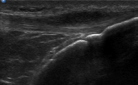
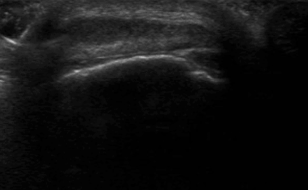
Ultrasound images at five weeks post trauma show a superficial hypo-echoic area in the medial-distal part of the patellar tendon. Focal areas with loss of collagen continuity.
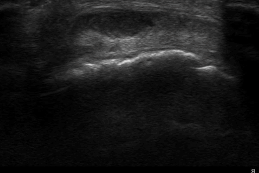
Ultrasound images at 4 1/2 months post trauma show an increasing reflectivity of the tendon and a return of the collagen bundle patterns in the longitudinal scan. Doppler imaging shows still one active vessel in the lesion. There is a complete return to sports at this stage.
0 Comments