Ultrasound in Rheumatology: Enthesitis on ultrasound
Dr Qasim Akram, Consultant Rheumatologist
Enthesopathy is described as an abnormally hypoechoic (loss of normal fibrillar architecture) and/or thickened tendon or ligament at its bony attachment (and may occasionally contain hyperechoic foci consisting of calcification) seen in 2 perpendicular planes that may exhibit doppler signal and/or bony changes including enthesophytes, erosions, or irregularity. It is classically seen with the spondyloarthropathies including psoriatic arthritis (peripheral spondyloarthrpathy) and ankylosing spondylitis (axial spondyloarthropathy).
Case Example
A 37-year-old male footballer presented with a 6/12 h/o pain and stiffness affecting his hands and feet with occasional episodes of swollen knees. He had a background history of psoriasis. He also complained of a tender and swollen right sided achilles tendon. Clinical examination revealed tender MCPs and MTPJs and a painful achilles tendon insertion site. Blood tests including immunology were normal. X rays were normal. A bedside ultrasound was performed .
Ultrasound findings
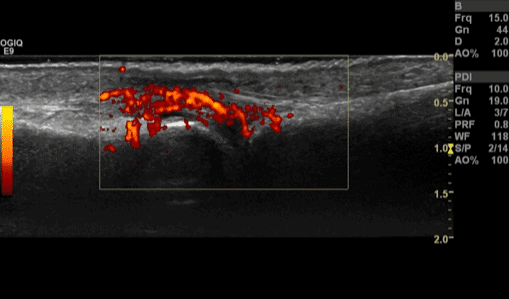
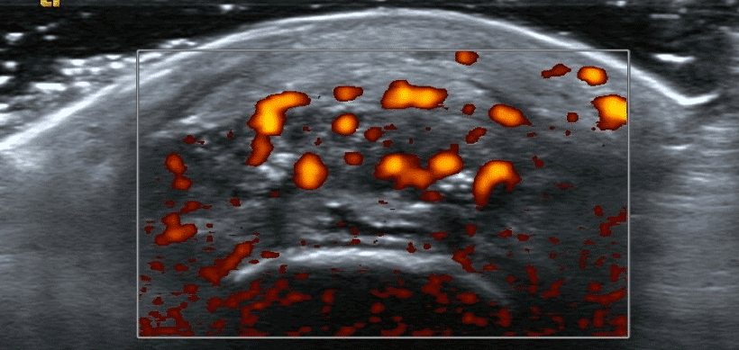
Ultrasound revealed synovitis of the MCPJs (Figure 1) (with intense doppler activity) and MTPJs (without any doppler activity). Achilles tendon insertion at the calcaneum (Figure 2 and 3) revealed evidence of enthesitis and enthesophytes with doppler activity.
Interpretation
He was classified as having a peripheral spondyloarthropathy ( and likely psoriatic arthritis) based on his clinical history, examination, and ultrasound findings and the absence of any auto-antibodies. The intense doppler activity on ultrasound and achilles tendon enthesitis was particularly pathognomonic.
References
- Wakefield et al. Musculo-skeletal ultrasound including definitions for ultrasonographic pathology. J Rheum 2005:32:2485-7.


0 Comments