Ultrasound in Rheumatology: Erosions on ultrasound
Dr Qasim Akram, Consultant Rheumatologist
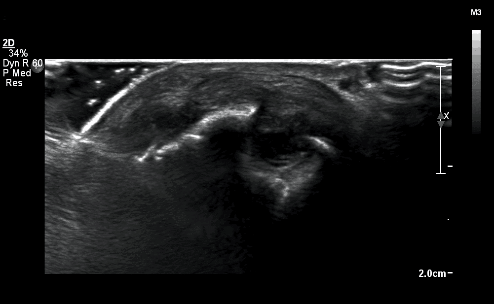
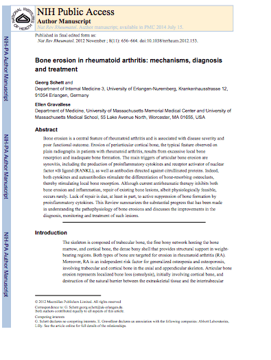
Erosions are described as an intra-articular discontinuity of the bony surface seen in 2 perpendicular planes. Ultrasound is up to seven times more sensitive at picking up these changes compared to x-rays thus very important in picking up signs of this to detect early disease. These tend to be present at accessible joint regions including the 2nd, 5th MCPJs and 1st and 5th MTPJs and radial heads and ulnar styloid surfaces.
Case Example
A man in their 40s presents with a 6/52 h/o pain and stiffness in his hands and feet which hasn’t responded to simple analgesics. Examination findings suggests pain in several of his joints in his hands and feet but no swelling. Blood tests are normal apart from a positive anti CCP antibody. X rays are normal.
Ultrasound findings
This demonstrated classical erosions present in the MCP joints.
Interpretation
Based on the fact that he was symptomatic, with evidence of clear erosive disease and ACPA positivity he was started on treatment for a seropositive rheumatoid arthritis.
References-
- Wakefield et al. Musculoskeletal ultrasound including definitions for ultrasonographic pathology. J Rheum 2005:32:2485-7.

0 Comments