Ultrasound Case Study
Flexor carpi radialis tendon sheath effusion, a red herring?
Clinical examination and history
This patient in their 60’s presented in clinic with swelling, pain and a feeling of tightness in the volar distal aspect of the forearm and wrist. There was no traumatic onset although she had noticed the onset of swelling a day or so after having sawed a piece of wood with a handsaw.
On palpation there was tenderness of the flexor carpi radialis (FCR) tendon and approximate distal insertion point. There was a visible swelling on the radial and volar aspect of the wrist. 1st CMCJ range of movement was preserved. Resisted 1st extensor compartment (Extensor pollicis brevis/ abductor pollicis longus) testing was normal. Wrist range was slightly restricted.
Ultrasound and other imaging appearances
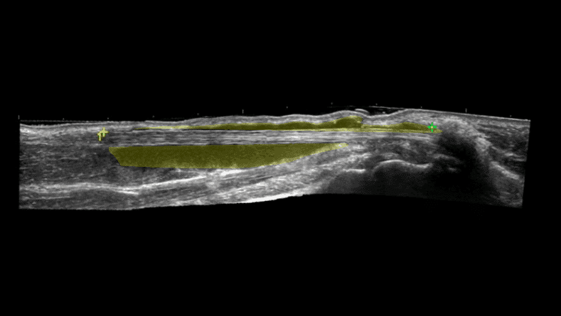
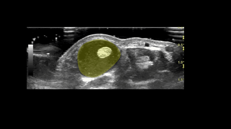
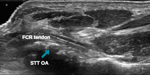
The ultrasound image encountered is as shown in Figure 1-3, a large effusion in the tendon sheath of the FCR. There was no hyperaemia on power doppler. Further evaluation of the distal part of the FCR showed marked osteophyte formation of the Scaphoid Trapezoid/Trapezium (STT) joint complex (Figure 3). The insertion of FCR on the 2nd Metacarpal however appeared intact.
Ultrasound report: ‘ The flexor carpi radialis tendon is intact, but there is a large effusion in the distal tendon sheath. The distal insertion is adjacent to a region of degenerative change of the STT articulation. Further X-ray may be helpful to evaluate further.’
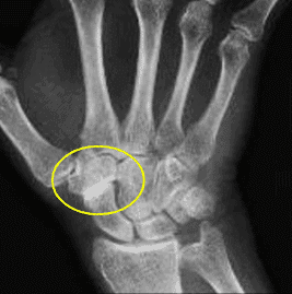
An X-ray was requested to clarify the STT articulation. The X-ray demonstrated advanced degenerative changes of the STT joint complex (Figure 4). The findings correlate well with tenosynovitis secondary to OA of the STT joint as described in a pictorial essay by Luong et al (2014).
The effusion within the distal FCR tendon sheath is often a reflection of STT pathology, and a red herring. The article below provides excellent anatomical and sonographic discussions around this point. It is not a free access paper therefore we cannot provide these for you.
Having modified her activities for many months and having tried exercises the situation did not improve. This lady was referred to the Orthopaedic hand specialist to explore further Orthopaedic management.
Learning points:
- Changes in the flexor carpi radialis are often a reflection of pathology of the STT articulation – make sure you check that and all other structures in this region including 1st CMCJ and 1st extensor compartment.
- Treatment focussed to the flexor carpi radialis may not be effective, and it may not need treatment.
Useful references for further reading:


0 Comments