Ultrasound Case Study
Freiberg disease of the 2nd MTPJ
Clinical examination and history
This patient presented with a two year history of forefoot pain. There was no history of trauma, or change in activity levels that preceded the onset of symptoms. Pain was localised to the region of the second and third MTPJs (Metatarsophalangeal joints). Movement testing of the 2nd MTPJ was uncomfortable but there was no significant laxity noted. As should be the case, differentiating the source of symptoms in this area is important and ultrasound can facilitate this.
Ultrasound and other imaging appearances
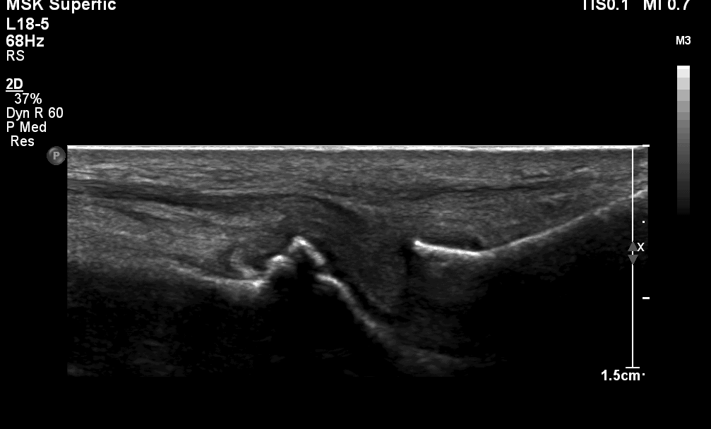
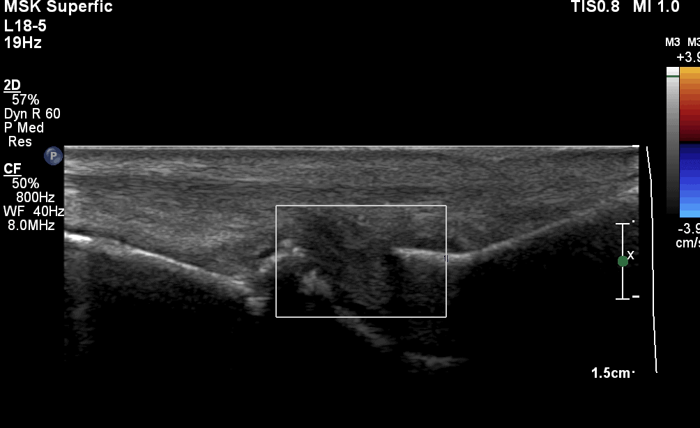
The ultrasound image encountered is demonstrated showing the characteristic appearance of an irregular deformed flattened metatarsal head in a longitudinal view (Figure 1). It is important to also evaluate the metatarsal shaft for periosteal reaction which can be present, although it was not in this case.There was no evidence of synovitis (Figure 2). The plantar plate was intact, and there were no bursa;/neuroma complexes in the 2/3 or 3/4 webspaces.
Ultrasound report ‘ The 2nd MTPJ demonstrated bony features suggestive of Freiberg disease. There was no evidence of synovitis. Further correlation with X-ray imaging is advised.’
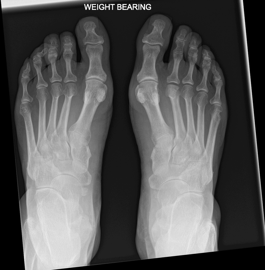
This can also be seen to correlate with the X-ray showing flattening of the right 2nd metatarsal head (Figure 3)
Pathology of Freiberg disease
Freiberg disease is avascular necrosis of the metatarsal head, most commonly involving the second, however it can present less commonly in others. The second metatarsal is affected in 68%, the third metatarsal in 27%, the fourth in 3%, and the fifth only rarely. The cause of Freiberg disease is often considered to be multi factorial. Two more likely causes are classified as trauma or vascular. The second metatarsal is the least mobile, thereby conferring the greatest stress at that metatarsal head distally (Cerato, 2011).Hallux rigidus, hallux valgus, or any malalignment that disrupts the normal weight-bearing function of the first ray similarly increases the load assumed by the second metatarsal predisposing to potential repetitive microtrauma (Cerato,2011). Also a vascular cause has been suggested as the second metatarsal is supplied by branches from the medial deep plantar artery and the dorsal metatarsal artery. Anatomic studies conducted have shown evidence of vascular variations, as well as absence of normal arterial flow to the affected metatarsal, rendering the blood flow to collateral circulation (Shane et al, 2013).
Learning points
- Be aware of the normal appearance of the metatarsal head on ultrasound and the distinctive appearance of the changes often associated with Freiberg disease.
- Consider further imaging if not previously performed.
- Evaluate the metatarsal shaft for periosteal changes as well as the metatarsal head.
- Always evaluate other structures in the forefoot to build a complete picture – including plantar plate, flexor tendon, intermetatarsal bursa/neuroma.
Useful references for further reading
Cerrato,RA (2011) Freiberg’s disease. Foot Ankle Clin 2011 Dec;16(4):647-58.


The only definite difference on XR is the increased joint space at 2nd mtpj on the affected side (right).
But very nice work and thank you for doing this service for learners like me.
AW