Starting out series – Article 1
How to orientate MSK ultrasound images
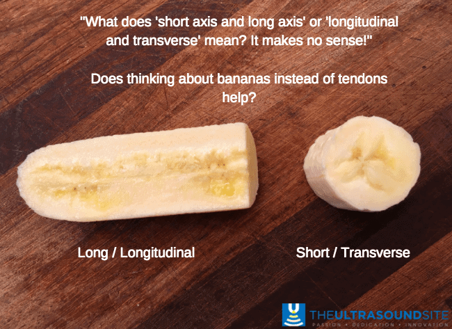
Sonographers often talk about their images in either the transverse or the longitudinal plane, or to confuse matters further..the short or long view. It is of fundamental importance that you understand what you are looking at and what this jargon means!
Longitudinal or long view
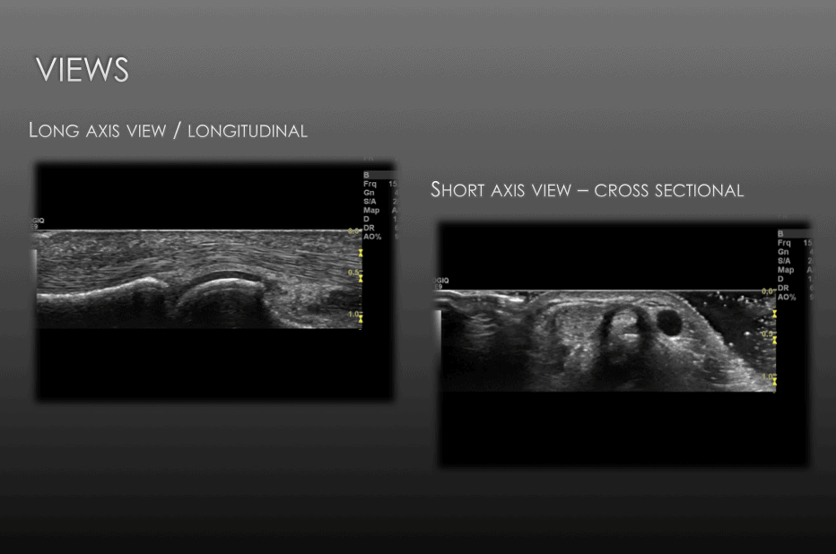
Lets start with the longitudinal view, which and may be termed a sagittal view in other forms of imaging. Below is a longitudinal view of a banana and also a tendon .
Transverse or short view
The transverse or short view..the easiest way to conquer this view is to think of a cross-sectional view looking up/down the structure.
So why do we bother to take both longitudinal and transverse views. Well we don’t always, and certainly with some MSK structures it is not common place to always interrogate with both a transverse and longitudinal view. For example, the common extensor tendon at the elbow is rarely viewed in a transverse view, and the reverse is true for the subscapularis which is not often viewed transversely. It is however good practice to look at both views to improve the accuracy of your images. We know that ultrasound is highly user dependent , and subtle changes in angulation of the probe can lead to very different conclusions and artefacts such as anisotropy. If you are therefore suspicious of for example a tear of a tendon it is good practice to convince yourself with both a longitudinal and transverse view.
As always, please comment and post your thoughts on this article or the topic in general!


beneficial thanks
Good and very light article for beginners. Please, send the next!!
Excelent interpretation of what happens to all of us at the beginning, Regards!
That is amazing that there are so many ways to look inside the human body and figure out what is wrong without having to operate. The way you used a banana for the analogy made things make so much more sense. It would be so fun to have the chance to use one of these machines and take a look at all the different kinds of muscles and tendons.
very good