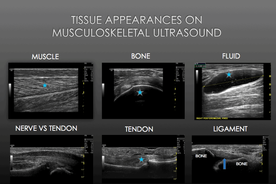
What do different tissues on ultrasound look like?
In the first of this series on the basics of using MSK ultrasound, we discussed how you can orientate yourself to the transverse/longitudinal image on ultrasound. I thought it would be useful to next discuss the appearance of different tissues on ultrasound. This is another key aspect to help orientation, as when you can appreciate the relationship of tendons to bones, and tie that together with your anatomical knowledge you can quickly develop a blueprint in your mind of where you are and what you are looking at.
I will cover the key tissues that you will encounter
Bone
Bone is represented as a very bright structure and appears ‘hyperechoic’. It creates a significant acoustic impedence mismatch and therefore is very reflective and shows as bright white (hyperechoic) on the image. No sound waves can pass through bone and therefore deep to it will always be dark.
Muscle
Muscle presents as hypoechoic, with some internal signals as a result of collagen fibres. The echotexture of normal skeletal muscles consists of a relatively dark or ‘hypoechoic’ background reflecting muscle fascicles along with linear hyperechoic strands related to fibroadipose septa (perimysium).
Tendon
Normal tendons as seen with the achilles images here, show tightly packed hyperechoic lines representing the fibrils of the tendon. On the transverse image the fibrillar pattern is presented as multiple hyperechoic dots in a tightly packed bundle.
Nerve
Ultrasound demonstrates nerves as ‘honeycomb’ or ‘pepper pot’ like structures composed of hypoechoic spots embedded in a hyperechoic background. They appear distinctly different to tendons in a transverse/short axis image as you can see here with the median nerve in the carpal tunnel.
Fluid
Fluid presents has an anechoic appearance on ultrasound, and can be confirmed with dynamic interrogation as it should respond to pressure. You can see here the anechoic or black appearance of fluid within the superficial infrapatellar bursa of the knee.
As always, please comment and post your thoughts on this article or the topic in general!

an excellent images overview-specially for beginners-thanks
thank you for that, would be even better if one could zoom in.