Shoulder Ultrasound
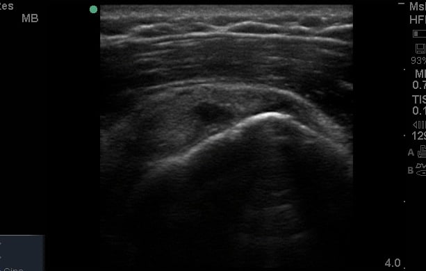
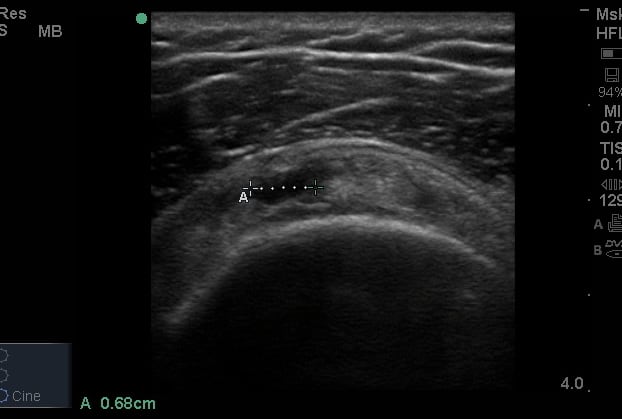
What structures can we see at the shoulder?
The shoulder is probably the most common area that you will be asked to ultrasound if you are performing diagnostic lists. It can however be quite challenging technically, due to the anatomy and bony outline of the humeral head. It is one of the more extensively researched regions of musculoskeletal ultrasound, and as always it is vitally important to establish the clinical significance of findings. In light of the fact that we know asymptomatic pathology is common in this region, particularly with increasing age, we should be critical of findings and relate them to the overall clinical picture.

'How to' tutorial videos for the shoulder
Case Studies
We have put together a series of case studies which describe how ultrasound was utilised alongside a clinical examination. Discussing how its use did or did not change the patient journey, alongside some useful references.
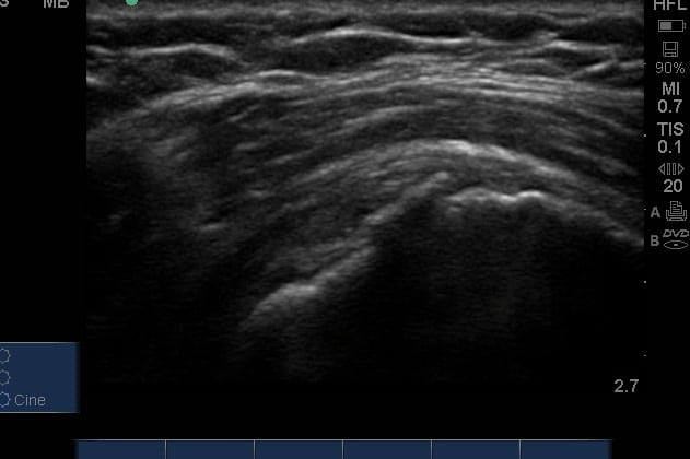
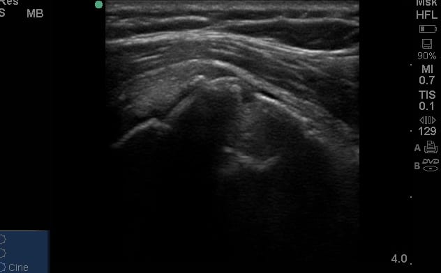
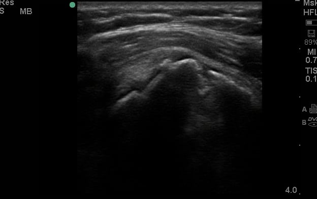
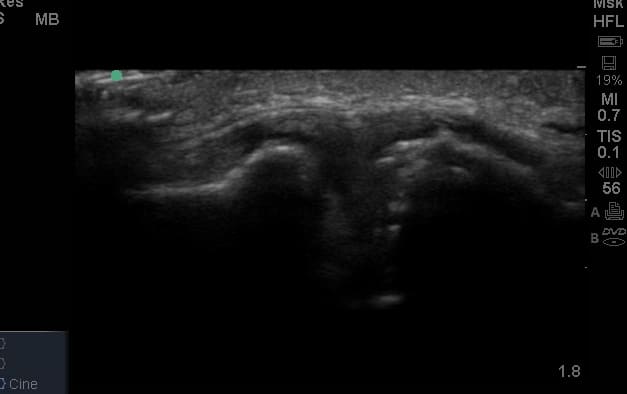
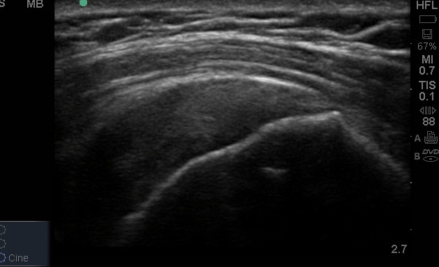
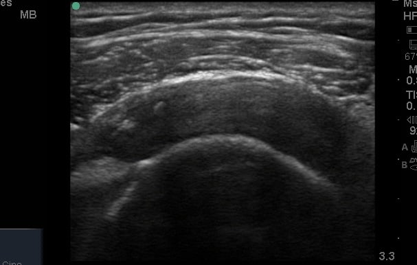
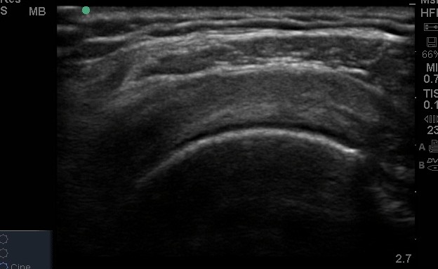
Gallery


Free access MSK ultrasound articles


Ultrasound pathology videos
Register to receive our newsletter including case studies
Anatomy

The shoulder joint, or glenohumeral joint, is a ball socket joint. It is composed of the flat glenoid fossa and the large round humeral head.With the glenoid being so flat, there is little bony congruity to joint which provides it with such mobility. the articular surfaces are covered by hyaline cartilage. A region of fibrocartilage, known as the labrum, deepens this joint to improve the stability.
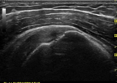
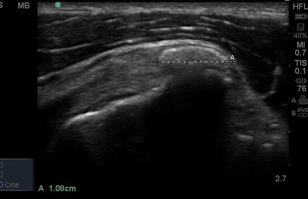
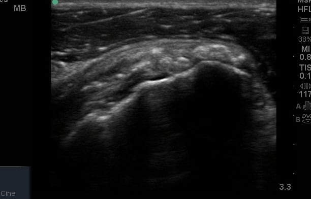
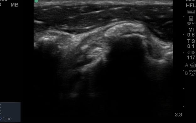
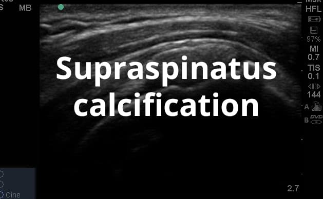
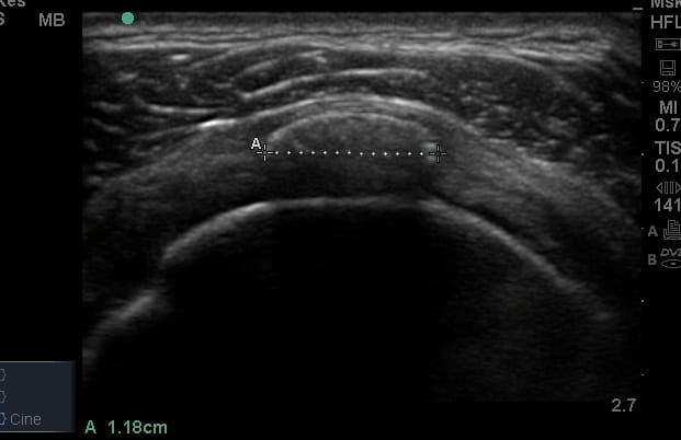
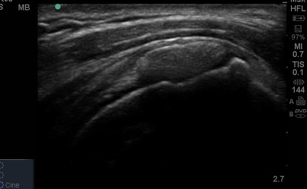
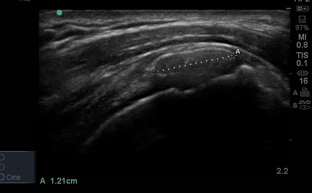
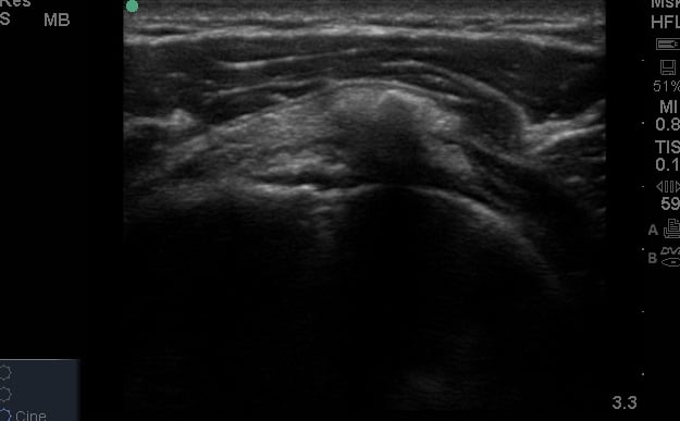
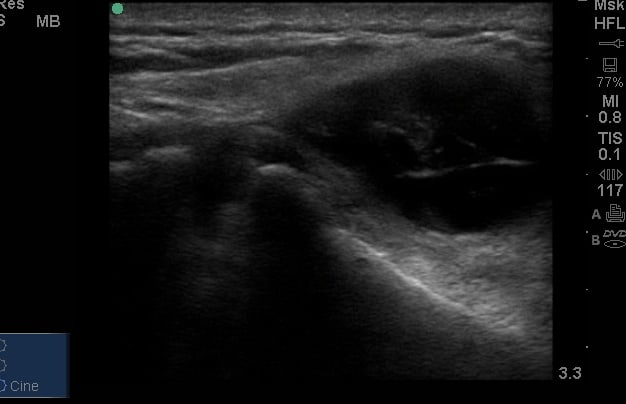
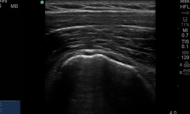
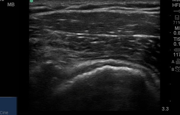
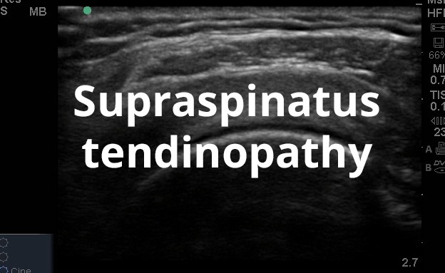
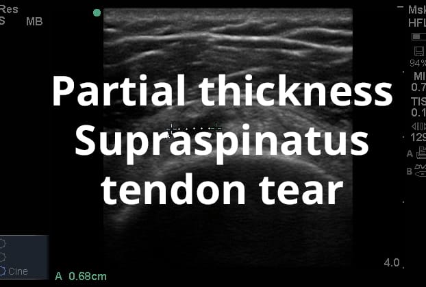
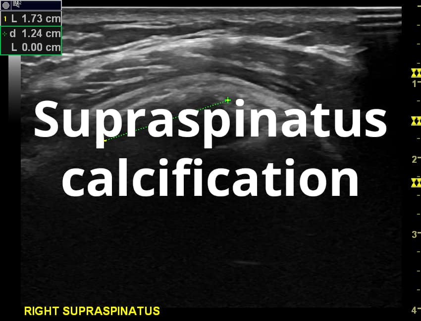
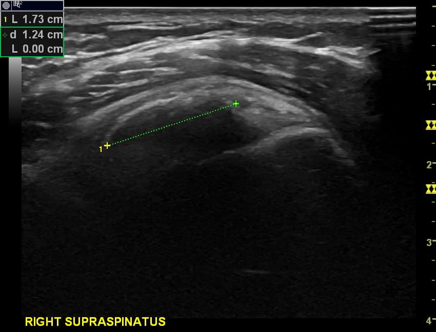
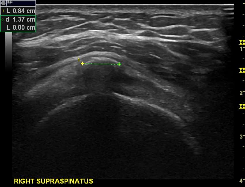
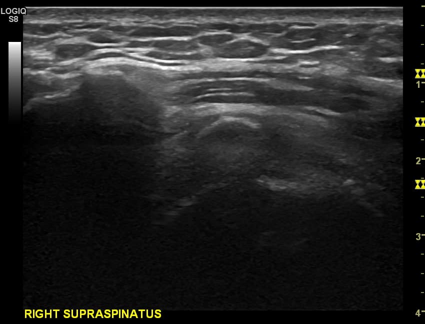
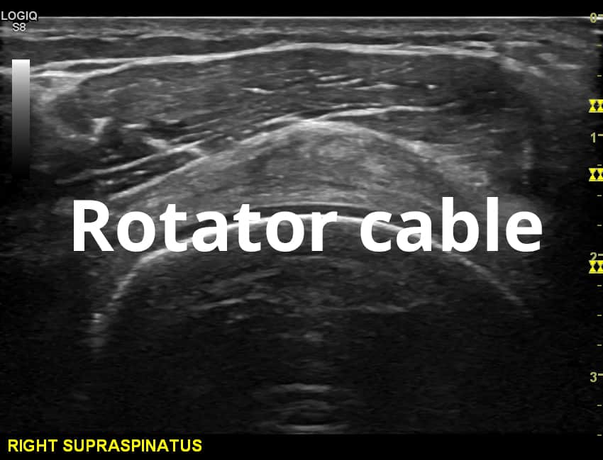
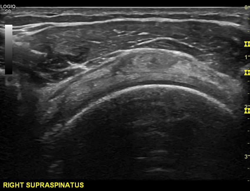
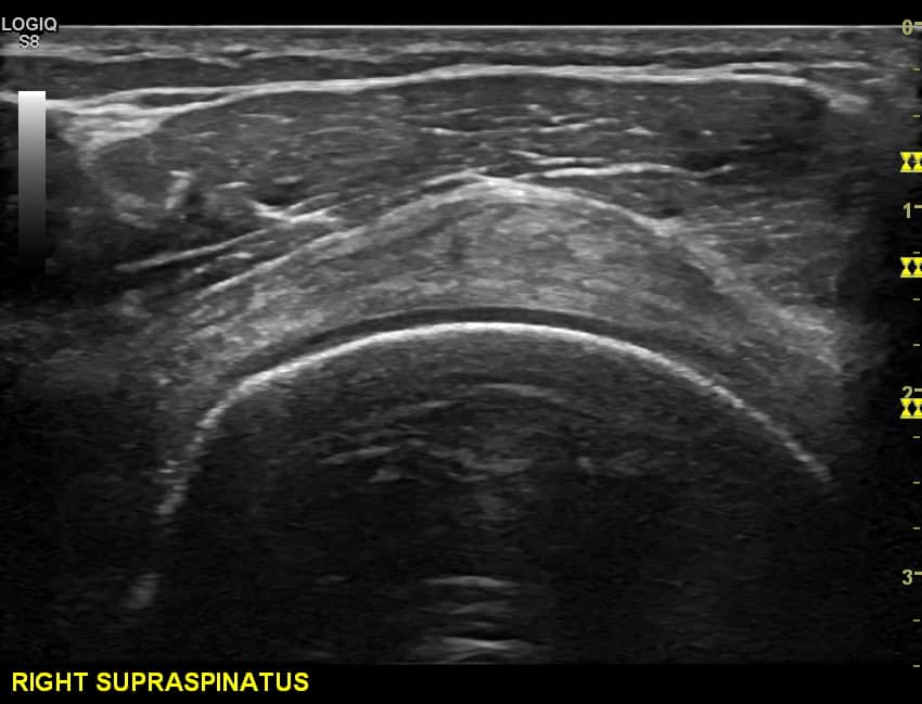
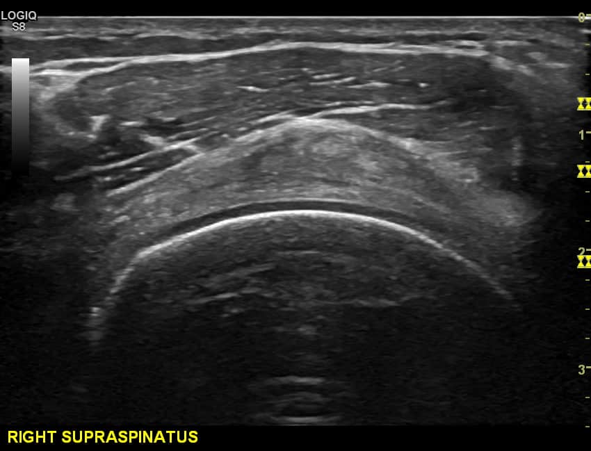
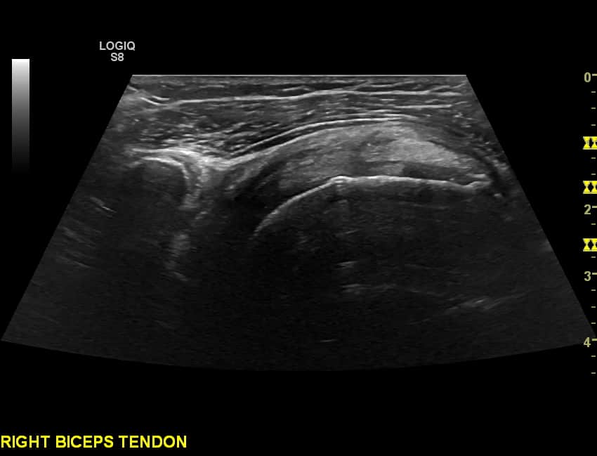
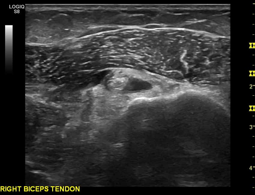
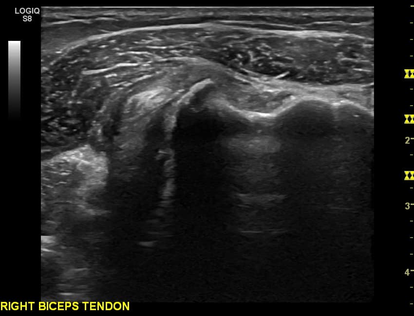
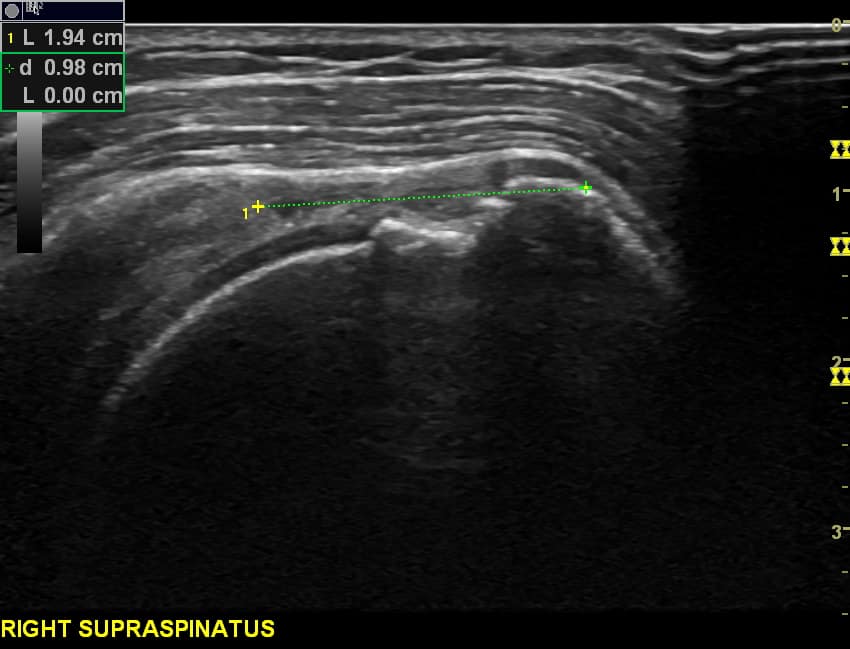
There are four rotator cuff muscles - The Subscapularis, Supraspinatus, Infraspinatus and Teres Minor. These will commonly be investigated with ultrasound. The Subscapualris and Supraspinatus are seperated by a space known as the rotator interval. This contains the long head of biceps tendon and the coracohumeral ligament. Along with the rotator cuff , the deltoid and teres major muscles are also present and help move the shoulder.
Anatomy articles of interest

