by Stuart Wildman | Oct 18, 2017
What do different tissues on ultrasound look like? In the first of this series on the basics of using MSK ultrasound, we discussed how you can orientate yourself to the transverse/longitudinal image on ultrasound. I thought it would be useful to next discuss the...
by Stuart Wildman | Sep 16, 2017
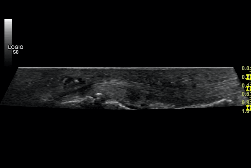
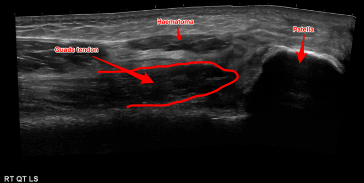
Ultrasound Case Study Quads tendon rupture Jon Sharpe, Consultant MSK Radiologist This patient had suffered a fall and developed sudden anterior knee pain and inability to straight leg raise. Interesting just how much you can see on the plain films and how closely it...
by Stuart Wildman | Sep 13, 2017
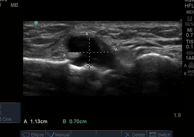
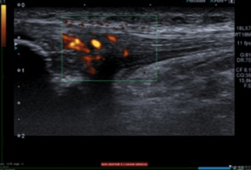
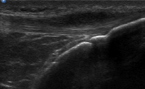
Ultrasound Case Study Pretibial ganglion following ACL reconstruction Stuart Wildman, Extended Scope Physiotherapist and MSK Sonographer This interesting case was referred for assessment after the patient had experienced 6 months of anterior knee pain, with a...
by Stuart Wildman | Sep 13, 2017
Ultrasound Case Study Patella tendinopathy and non-union of the ossification centres Dr Matthieu Sailly (@MedecinsDuSport) Sports Medicine Physician, Switzerland Following on from the recent case I posted on patellar tendinopathy and the role of diagostic MSK...
by Stuart Wildman | Sep 13, 2017
Ultrasound Case Study Acute patella tendon impact injury Niek Vink, Physiotherapist and MSK Sonographer((Dutch National Training Centre for Ultrasound, http://www.nt-e.nl/) This interesting case gives insight into the sonographic appearance of an acute patella tendon...