Ultrasound Case Study
Finger flexor tenosynovitis?
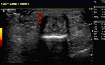
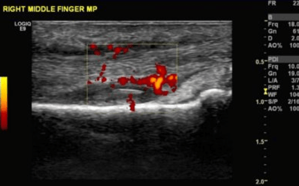
Initial images ultrasound images show thickening of common flexor tendon of the finger associated with hyperaemia on power Doppler suggestive of tenosynovitis.
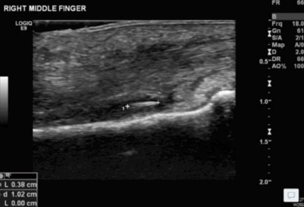
I was concluding the scan, and the patient suddenly recalled that she did some work in her garden a few weeks earlier and since then she had noticed symptoms. She also reported the sensation that something was inside. Following a further careful examination under ultrasound there was indeed a focal hyperechogenic lesion noted underlying the flexor tendon over the middle phalanx associated with minimal granulation tissue. Appearances are consistent with a foreign body, potentially a wood splinter. The patient was referred back to the Orthopaedic team who surgically removed a wood splinter.
Ultrasound report
There is a focal hyper echogenic lesion measuring 4mm in length noted underneath the flexor tendons near to the PIP joint of the right middle finger associated with a moderate degree of thickening of common flexor tendon sheath and neovascularity on power doppler. Appearances are consistent with a foreign body possible splinter within the common flexor tendon sheath associated with tenosynovitis.
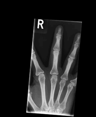
Subsequent xray reports ‘No radiopaque foreign body / periosteal reaction is seen’ , the images can be seen below.
The lack of posterior shadowing is suggestive of a less dense structure compared to a needle or metal. This can also be seen in another case study of a foreign body at the ankle.
Some articles on foreign bodies and their sonographic appearance.
We hope you have found this case study of interest.
Feel free to comment in the feedback section at the bottom of this page as well. Your support is what helps the website grow and encourage more case studies to be posted.
If you would like to post a case study please contact us at the email below!
Feedback is always appreciated [email protected]



0 Comments