Ultrasound in Rheumatology: Synovitis and ultrasound
Dr Qasim Akram, Consultant Rheumatologist
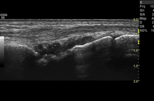
Synovitis or synovial hypertrophy is described as being an abnormal hypoechoic, poorly compressible and non-displaceable intra-articular tissue which may exhibit doppler signal. In contrast, synovial effusion is described as being an abnormal anechoic, compressible and displaceable intra-articular material that does not exhibit doppler signal.
Synovitis can be graded according to a scale of 0-3. Grade 0 means that there is no synovial thickening. Grade 1 means there is minimal synovial thickening without bulging over the tops of the bones. This can be seen in normal patients who do not have disease pathology. Grade 2 means there is more extensive synovial thickening with bulging over the tops of the periarticular bones. Grade 3 is more extensive thickening with extension beyond the joint.
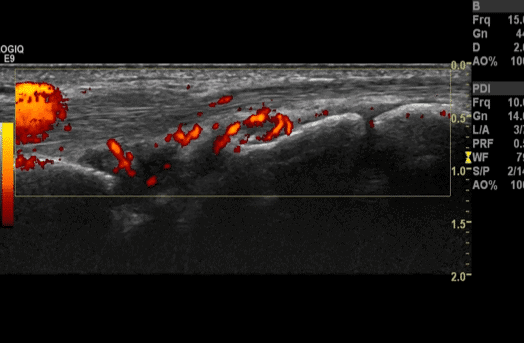
Doppler activity can also be graded. Grade 0 means there is no doppler activity. Grade 1 means there is some activity, Grade 2 means less than <50% area of the joint and Grade 3 is >50% of the area of the joint.
In essence this means that the greater the degree of grey scale synovitis and doppler grading the more extensive the disease.
Case example
A patient presents to the early inflammatory arthritis clinic with a 4/52 h/o bilateral wrist and small joint pain and stiffness. The rest of her joints are unremarkable. She denies any other history. Clinical examination shows tender wrists and small joints but no underlying swelling. Blood tests show a positive rheumatoid factor and modestly elevated inflammatory markers. X rays are normal.
US findings
USS of her hands and wrists, performed at the bedside, demonstrated extensive wrist synovitis with corresponding doppler activity (Grade 3 synovitis and Grade 3 Doppler) and PIPJ synovitis:
Interpretation
Based on her history, physical examination findings, raised inflammatory markers and ultrasound scan showing synovitis, a diagnosis of early rheumatoid arthritis was made. Treatment with corticosteroids and DMARDs was initiated immediately in line with best practice.
References-
- Wakefield et al. Musculo-skeletal ultrasound including definitions for ultrasonographic pathology. J Rheum 2005:32:2485-7.
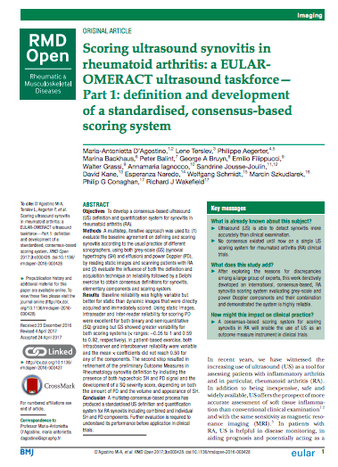
- D’ Agostini et al. Scoring ultrasound synovitis in rheumatoid arthritis- a EULAR-OMERACT ultrasound taskforce-Part 1: definition and development of a standardised consensus-based scoring system. RMD Open 2017 Jul 11;3(1): e000428. doi: 10.1136/rmdopen-2016-000428. eCollection 2017.
0 Comments