Ultrasound in Rheumatology: Tenosynovitis
Dr Qasim Akram, Consultant Rheumatologist
Tenosynovitis is described as being an abnormal hypoechoic/anechoic thickened tissue in the tendon sheath with or without doppler activity. It can affect both flexor and extensor tendons in rheumatic disease. Doppler activity tends to correspond to disease activity. When tendons that don’t have a tendon sheath are affected this is labelled as a paratenonitis. For example, Achilles tendon and extensor digitorum tendons at the level of the metacarpo-phalangeal joints. In rheumatic disease, early predictors of disease can be involvement of the digital flexor tendons as well as extensor tendons especially the extensor carpi ulnaris (ECU) tendon.
Case Example
A joiner presented with a 3/12 h/o wrist and pain located in the palms of his hands and fingers. Clinical examination revealed tenderness in his wrists including positive Finkelstein’s test for De Quervain’s tenosynovitis. His blood tests were positive for Rheumatoid Factor. X rays were normal.
Ultrasound findings
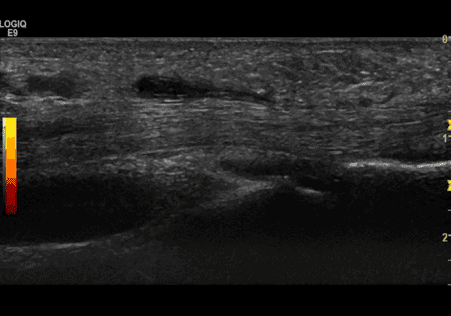
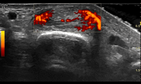
Ultrasound showed tenosynovitis of the extensor tendons (compartments 1&2) , index, middle finger flexor tendons and index finger extensor tendon.
Interpretation
Based on his history, examination, blood tests and ultrasound findings he was classified as having a seropositive rheumatoid arthritis. Immediate steroid treatment was initiated with DMARD initiation. He was referred for OT.
References-
- Wakefield et al. Musculo-skeletal ultrasound including definitions for ultrasonographic pathology. J Rheum 2005:32:2485-7.
- Sahbudin et al. The role of ultrasound defined tenosynovitis in the prediction of rheumatoid arthritis development. Rheumatology 2018:57: 1243-1252.
- Tenosynovitis of the extensor carpi ulnaris predicts erosive progression in early rheumatoid arthritis. Ann Rheum Dis 2011: 70: 2049-2050.

0 Comments