by Stuart Wildman | Oct 18, 2017
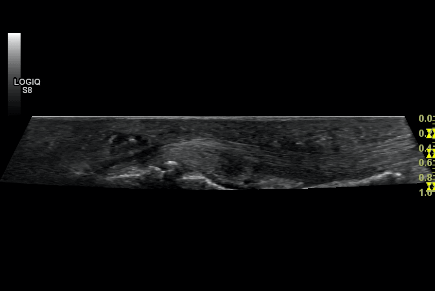
What do different tissues on ultrasound look like? In the first of this series on the basics of using MSK ultrasound, we discussed how you can orientate yourself to the transverse/longitudinal image on ultrasound. I thought it would be useful to next discuss the...
by Stuart Wildman | Sep 25, 2017
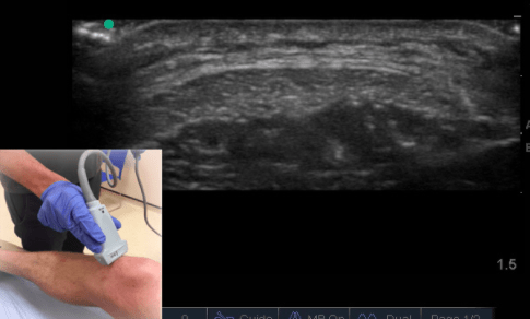
Ultrasound Case Study Anisotropy, friend or foe? David Baker, Extended Scope Physiotherapist and MSK Sonographer In order for the ultrasound machine to create an image the systems software must make a number of assumptions. The assumptions that the machine makes are:-...
by Stuart Wildman | Sep 17, 2017
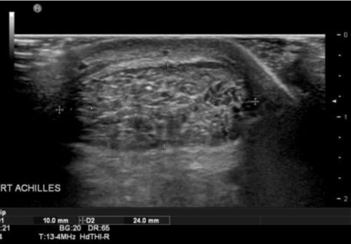
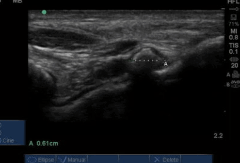
Ultrasound Case Study Achilles tendinopathy Peter Gettings, Physiotherapist and MSK Sonographer This male patient in their early 60’s was referred to our ultrasound clinic for evaluation of his bilateral Achilles pain. He had been assessed by a colleague and...
by Stuart Wildman | Sep 17, 2017
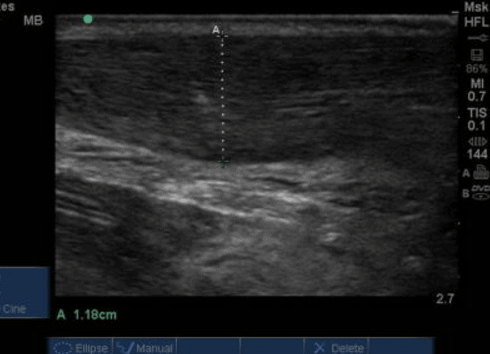
Ultrasound Case Study Fluoroquinolone induced achilles tendinopathy This case study relates to a patient in his late 60’s, who was started on Ciprofloxacin a few years earlier for a suspected infection. After taking for five days he spontaneously experienced...
by Stuart Wildman | Sep 17, 2017
Ultrasound Case Study Loose body of the ankle joint Stuart Wildman, Extended Scope Physiotherapist and MSK Sonographer This patient in their 30’s attended clinic complaining of several years of difficulties with his right ankle. He was previously a keen...