Ultrasound in Rheumatology: Rheumatoid and Ultrasound
Dr Qasim Akram, Consultant Rheumatologist
Rheumatoid arthritis is a systemic inflammatory disease characterised by the presence of a destructive polyarthritis affecting the small joints of the hands and feet and wrists (although can affect any synovial joint). If left untreated inflammation and joint destruction leads to loss of physical function, inability to carry out activities of living and maintaining employment.
Commonly affects adults although can occur at younger ages. Females tend to be more affected more than males
In terms of systemic disease, other internal organs that are affected include the lungs, kidneys, eyes and cardiovascular system.
Clinical Presentation
Clinical features include symmetrical synovitis (manifested as pain, swelling or stiffness) of the small joints of the hands and feet with sparing of the DIPJs. Tenosynovitis and less commonly bursitis may also be present. In early disease, can present as a monoarthritis or in asymmetrical distribution. Early morning stiffness is a hallmark of the disease and lasts for longer than 30 minutes.
Common joints involved include hands, wrists, elbows, shoulders and knees followed by hips and TMJs less commonly.
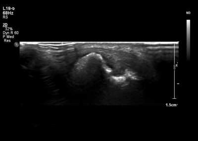
Diagnosis is based on clinical and laboratory tests. EULAR/ACR 2010 criteria is often used to classify the disease. RhF is present in 70-80% of all patients with RA. However, its specificity is poor and can be elevated in several other disease states. Antibodies to CCP have a similar sensitivity but a much higher specificity profile (>90%). Inflammatory markers such as CRP and ESR are usually elevated and can indicated severity of disease. Synovial fluid analysis can reveal an inflammatory effusion.
Pathophysiology
Pathophysiology of this condition is due to a complex interplay of environmental, genetic and infective triggers. Usually, this results in an immune response (involving B cells, T cells and cytokines) occurring within joints and tendons causing synovitis, tenosynovitis and erosive disease.
Typical US findings
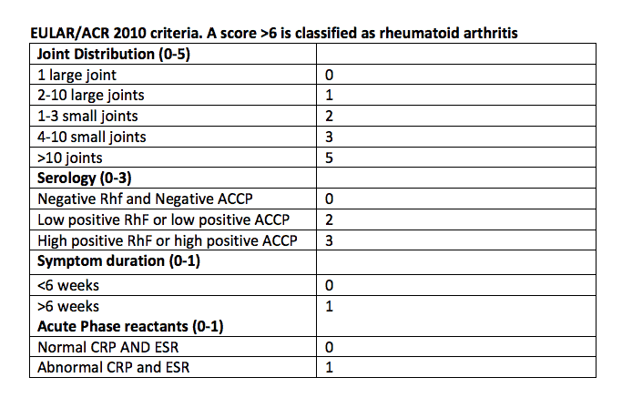
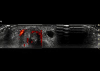
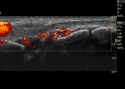
Typical ultrasound features include synovitis/synovial hypertrophy (with doppler activity), effusions, erosions and tenosynovitis. The extent of synovitis and doppler activity can be a measure of disease severity.
Typical sites include ECU, FPL and flexor tendons. Others include index, middle MCP and PIPJs, ulnar styloid, and 2nd, 3rd and 5th MTPJs.
See the links below to specific features that are often evident in Rheumatoid arthritis
References:
Clunie G et al. Oxford Handbook of Rheumatology. 4th Oxford University Press 2018.
0 Comments